Inflammatory Arthritides


Inflammatory Arthritides


The inflammatory arthritides comprise a group of different and for the most part systemic disorders (see Fig. 12.1) that have in common one important feature: inflammatory pannus eroding articular cartilage and bone (Fig. 14.1). An overview of the clinical and radiographic hallmarks of the various inflammatory arthritides is shown in Table 14.1.


Erosive Osteoarthritis

Erosive osteoarthritis was first described by Kellgren and Moore in 1952 and reintroduced in 1961 by Crain, who called it interphalangeal osteoarthritis. He defined this disorder as a localized variant of osteoarthritis involving the finger joints, characterized by degenerative changes with intermittent inflammatory episodes leading to deformities and ankylosis. In 1966, Peter and Pearson coined the term erosive osteoarthritis, and Ehrlich in 1972 described it as inflammatory osteoarthritis, based on the clinical symptoms of swelling, tenderness, erythema, and warmth. It can be defined as a progressive disorder of the interphalangeal joints with severe synovitis, superimposed on the changes of degenerative joint disease. Although the cause is still unclear, several investigators have suggested hormonal influences, metabolic background, autoimmunity, and heredity as being involved.

Erosive osteoarthritis is a progressive inflammatory arthritis seen predominantly in middle-aged women. Only rarely are men affected, with an estimated female-to-male ratio of 12:1. Patients’ ages range from 36 to 83 years, and mean age of onset is 50.5 years. This condition combines certain clinical manifestations of rheumatoid arthritis with certain imaging features of degenerative joint disease. Involvement is limited to the hands, with the proximal and distal interphalangeal joints being the most frequently affected. Large joints, such as the hip or shoulder, are only rarely involved. The arthritis usually begins abruptly and is characterized by pain, swelling, and tenderness of the small joints of the hands. Also described are throbbing paresthesias of the fingertips and morning stiffness.

In the early stage of the disease, the main feature is symmetric synovitis of the interphalangeal joints. Later, this is followed by articular erosions, which exhibit a characteristic radiographic feature named the “gull-wing” deformity by Martel. This configuration is seen as a result of central erosion and marginal proliferation of bone (Fig. 14.2); Heberden nodes may also be present. Periosteal reaction taking the form of linear or fluffy bone apposition over the cortex near the affected joints is occasionally observed. Swelling of soft tissue, usually fusiform, may be present around involved articulations (Fig. 14.2C); however, periarticular osteoporosis is rarely present. Later in the disease process, bone ankylosis of the phalanges may develop. Approximately 15% of patients with erosive osteoarthritis may have clinical, laboratory, and radiographic manifestations of rheumatoid arthritis (Fig. 14.3). The exact relationship between these two conditions is still unclear. Some investigators believe that erosive osteoarthritis is actually rheumatoid arthritis originating in unusual sites but subsequently progressing to the articulations that are more typically involved. Others suggest that each is a distinct entity, citing as evidence the fact that the synovial fluid of patients with rheumatoid arthritis does not resemble that of patients with erosive osteoarthritis, that the immunologic abnormalities commonly seen in rheumatoid arthritis are absent in the latter condition, and that the serologic test for rheumatoid factor is negative.

Occasionally, a variant of erosive osteoarthritis may be seen as one of the features of Cronkhite-Canada syndrome. This rare systemic disorder also manifests with generalized gastrointestinal polyposis, hyperpigmentation of the skin, and nail atrophy.


Treatment

The main objective of therapy in patients with inflammatory erosive osteoarthritis is relief of pain and restoration of joint function. Nonpharmacologic therapy includes physical and occupational therapy. Range-of-motion exercises and moist heat, in the form of a paraffin bath, are helpful. Pharmacologic methods include analgesics, nonsteroidal antiinflammatory drugs (NSAIDs), and corticosteroids. Selected cases have also been treated with methotrexate and oral gold salts. Recently, promising results have been achieved with administration of hydroxychloroquine in patients who did not respond to NSAIDs. Also good results have been reported after subcutaneous injections of adalimumab, and intraarticular injections of infliximab. Surgical intervention is often necessary for the relief of persistent pain and the correction of severe deformities. One of the most effective procedures is joint replacement by means of silicone-rubber arthroplasties (Fig. 14.3B). The indications for this type of surgery are loss of the joint space, synovial proliferation with joint destruction, loss of normal alignment, and uncontrollable pain.


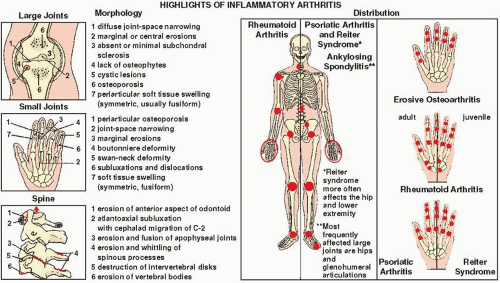
FIGURE 14.1 Inflammatory arthritides. Highlights of the morphology and distribution of arthritic lesions in the inflammatory arthritides.


Rheumatoid Arthritis


Adult Rheumatoid Arthritis

Rheumatoid arthritis is a progressive, chronic, systemic inflammatory disease affecting primarily the synovial joints; women are affected three times more often than men. The course of the disease varies from patient to patient, and there is a striking tendency toward spontaneous remissions and exacerbations. The detection of rheumatoid factor, representing specific antibodies in the patient’s serum, is an important diagnostic finding. Although it is still debatable, some investigators also include under this rubric a condition called seronegative rheumatoid arthritis (see later), in which patients present without rheumatoid factor but with the clinical and radiographic picture of rheumatoid arthritis.


Rheumatoid Factors

Rheumatoid factors, so widely used by clinicians, are antigammaglobulin antibodies that are elaborated in part by rheumatoid synovium. Rheumatoid factors in synovial fluid are either of the IgG or of the IgM variety. They combine with their antigens (immunoglobulin G [IgG]) to form immune complexes. These complexes activate the complement system, which releases mediators responsive for producing inflammation within the joint structures. Because rheumatoid factors can be found in the joint fluids of patients with nonrheumatoid disorders, their presence alone is not diagnostic of rheumatoid arthritis. However, finding high titers of these factors in a joint effusion strongly suggests the diagnosis of rheumatoid arthritis. Early in the course of disease, rheumatoid factors may be demonstrated in the synovial fluid before they are positive in the serum, allowing early diagnosis.

Rheumatoid factors participate in the pathogenesis of rheumatoid arthritis through the formation of local and circulating antigen-antibody complexes. In synovial fluid, IgM and IgG rheumatoid factors can combine with antigen (IgG) to form immune complexes. The complement system is activated, resulting in the attraction of polymorphonuclear leukocytes into the joint space. Discharge of their hydrolytic enzymes causes the destruction of joint tissues. The process initiating these events is as yet unknown.

Rheumatoid factors are not, however, absolutely diagnostic of rheumatoid arthritis and are found in the synovial fluid and serum in approximately 70% to 80% of patients with a clinical diagnosis of rheumatoid arthritis. In rheumatoid arthritis of recent onset, the test for rheumatoid factors initially may be negative in serum or synovial fluid, but later may become positive. Patients who are seropositive at the onset of their disease will often sustain persistent disease activity and disability. Patients with rheumatoid arthritis with subcutaneous nodules almost always will have positive agglutination tests, generally in high titer.


Imaging Features

Rheumatoid arthritis is characterized by a diffuse, usually multicompartmental, symmetric narrowing of the joint space associated with marginal or central erosions, periarticular osteoporosis, and periarticular soft-tissue swelling; subchondral sclerosis is minimal or absent and formation of osteophytes is lacking.


Large Joint Involvement

Any of the large weight-bearing and nonweight-bearing joints can be affected by rheumatoid arthritis. Regardless of the size of the joint and the site of involvement, certain imaging features can be identified that are characteristic of this inflammatory process.

Osteoporosis. In rheumatoid arthritis, unlike osteoarthritis, osteoporosis is a striking feature. In the early stage of the disease, osteoporosis is localized to periarticular areas, but with progression of the condition a generalized osteoporosis can be observed.

Joint Space Narrowing. This is usually a symmetric process with concentric narrowing of the joint. In the knee, all three joint compartments are involved (Fig. 14.4). Concentric narrowing in the hip joint leads to axial migration of the femoral head, which in more advanced stages may result in acetabular protrusio (Fig. 14.5). Cephalad migration of the humeral head may also be seen secondary to destructive changes in the shoulder joint and rupture of the rotator cuff (Fig. 14.6);
resorption of the distal end of the clavicle, which assumes a pencil-like appearance, may also be observed. Tear of the rotator cuff in this condition (Fig. 14.7) must be differentiated from the chronic traumatic form of this abnormality (see Fig. 5.62).


TABLE 14.1 Clinical and Imaging Hallmarks of Inflammatory Arthritides


Type of Arthritis


Site


Crucial Abnormalities


Technique*/Projection


Erosive Osteoarthritis (F; middle age)


Hands


Involvement of Proximal interphalangeal joints Distal interphalangeal joints


Gull-wing deformities associated with erosions


Heberden nodes


Joint ankylosis


Dorsovolar view


Rheumatoid Arthritis (F > M; presence of rheumatoid factor and DRW4)


Hands and wrists


Involvement of Metacarpophalangeal joints Proximal interphalangeal joints


Central and marginal erosions


Periarticular osteoporosis


Joint deformities: swan-neck, boutonnière, main-en-lorgnette, hitchhiker’s thumb


Dorsovolar view


Synovitis


Dorsovolar and lateral views


Hip


Narrowing of joint space


Erosions


Acetabular protrusion


MRI


Anteroposterior and lateral views


Knee


Narrowing of joint space


Erosions


Synovial cysts


Anteroposterior and lateral views


Ankle and foot


Involvement of subtalar joint


MRI


Anteroposterior and lateral views


Erosions of calcaneus


Lateral and Broden views


Lateral view (heel)


Juvenile Rheumatoid Arthritis


Hands


Joint ankylosis


Periosteal reaction


Growth abnormalities


Dorsovolar view (wrist and hand)


Knees


Growth abnormalities


Anteroposterior and lateral views


Cervical spine


Fusion of apophyseal joints


Anteroposterior, lateral, and oblique views


Rheumatoid Variants Ankylosing Spondylitis (M > F; young adult; 95% positive for HLA-B27)


Spine


C1-2 subluxation


Squaring of vertebral bodies


Syndesmophytes


“Bamboo” spine


Paravertebral ossifications


Lateral view in flexion


Anteroposterior and lateral views


Sacroiliac joints


Inflammatory changes


Fusion


Posteroanterior and Ferguson views


Pelvis


Whiskering of iliac crests and ischial tuberosity


Anteroposterior view


Reiter Syndrome (M > F)


Foot


Involvement of great toe articulations


Erosions of calcaneus


Anteroposterior and lateral views


Spine


Single, coarse syndesmophyte


Anteroposterior and lateral views


Sacroiliac joints


Unilateral or bilateral but asymmetric involvement


Posteroanterior and Ferguson views


Computed tomography


Psoriatic Arthritis (M ≥ F; skin changes; HLA-B27 positive)


Hands


Involvement of distal interphalangeal joints


Erosion of terminal tufts


Mouse-ear erosions


Pencil-in-cup deformities


Sausage digit


Joint ankylosis


Fluffy periosteal reaction


Dorsovolar view


Foot


Involvement of distal interphalangeal joints


Erosions of terminal tufts and calcaneus


Anteroposterior and lateral views (ankle and foot)


Spine


Single, coarse syndesmophyte


Anteroposterior and lateral views


Sacroiliac joints


Unilateral or bilateral but asymmetric involvement


Posteroanterior and Ferguson views


Enteropathic Arthropathies


Sacroiliac joints


Symmetric involvement


Posteroanterior and Ferguson views


Computed tomography


* Radionuclide bone scan is used to determine the distribution of arthritic lesions in the skeleton.


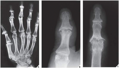
FIGURE 14.2 Erosive osteoarthritis. (A) Dorsovolar film of the left hand of a 48-year-old woman with erosive osteoarthritis shows the typical involvement of the proximal and distal interphalangeal joints. Note the “gull-wing” pattern of articular erosion, a configuration resulting from peripheral bone erosion in the distal side of the joint and central erosion in the proximal side of the joint associated with marginal bone proliferation. (B) Dorsovolar radiograph of the left thumb of a 51-year-old woman shows characteristic gull-wing erosion of the interphalangeal joint. Note adjacent fusiform soft-tissue swelling and lack of periarticular osteoporosis. (C) In another patient, a 50-year-old woman, gull-wing erosion is accompanied by periosteal reaction and fusiform soft-tissue swelling, very similar to psoriatic arthritis.


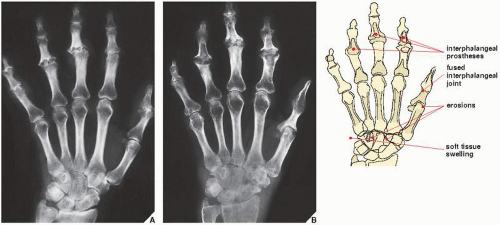
FIGURE 14.3 Progression of erosive osteoarthritis into rheumatoid arthritis. (A) Dorsovolar radiograph of the hand of a 58-year-old woman demonstrates the gull-wing configuration of erosive changes in the proximal interphalangeal joints and the distal interphalangeal joint of the small finger. Because of protracted pain and lack of response to conservative treatment, she underwent joint resection followed by implantation of silicone-rubber prostheses in the proximal interphalangeal joints of the index, middle, and ring fingers, together with fusion of the interphalangeal joint of the thumb and the distal interphalangeal joint of the small finger. Five years after surgery, the classic radiographic features of rheumatoid arthritis developed, involving the wrists (B), elbows, shoulders, hips, and cervical spine. Note the surgical fusion of interphalangeal joints of the thumb and fifth finger, as well as the spontaneous fusion of the distal interphalangeal joints of the index and ring fingers.


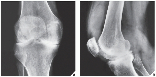
FIGURE 14.4 Rheumatoid arthritis. Anteroposterior (A) and lateral (B) radiographs of the knee of a 52-year-old woman with rheumatoid arthritis affecting several joints show tricompartmental involvement. Note the periarticular osteoporosis, joint effusion, and lack of osteophytosis.


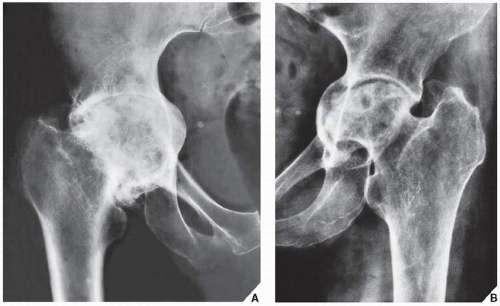
FIGURE 14.5 Rheumatoid arthritis. (A) Anteroposterior radiograph of the right hip of a 60-year-old woman with advanced rheumatoid arthritis shows concentric joint space narrowing, with axial migration of the femoral head leading to acetabular protrusion. Some superimposed secondary osteoarthritic changes are also present. (B) Anteroposterior radiograph of the left hip of a 64-year-old woman shows erosions of the femoral head and acetabulum, concentric narrowing of the hip joint, and acetabular protrusion.


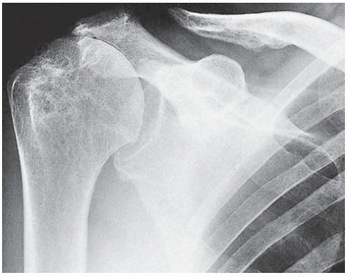
FIGURE 14.6 Rheumatoid arthritis. Anteroposterior radiograph of the right shoulder of a 72-year-old man with advanced rheumatoid arthritis shows upward migration of the humeral head secondary to rotator cuff tear, a common complication of rheumatoid changes in the shoulder joint. Note the characteristic tapered erosion of the distal end of the clavicle, erosions of the humeral head, and the substantial degree of periarticular osteoporosis.


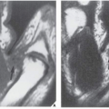
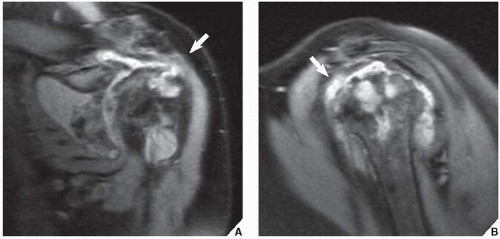
FIGURE 14.7 MRI of rheumatoid arthritis. (A) Oblique coronal and (B) sagittal proton density-weighted fat-suppressed MR images of the left shoulder of a 64-year-old woman show large articular and periarticular erosions, joint space narrowing, joint effusion, and a tear of the supraspinatus tendon (arrows), all the features of advanced rheumatoid arthritis.

Articular Erosions. Erosive destruction of a joint may be central or peripheral in location. As a rule, reparative processes are absent or very minimal; thus, there is no evidence of subchondral sclerosis or osteophytosis (Fig. 14.8), which may be present only if secondary degenerative changes are superimposed on the underlying inflammatory process (see Fig. 13.5).

Synovial Cysts and Pseudocysts. These radiolucent defects are usually seen in close proximity to the joint (Fig. 14.9). They may or may not communicate with the joint space.

Joint Effusion. Fluid can be best demonstrated in the knee joint on the lateral projection (see Fig. 14.4B). Fluid in the other large joints such as the shoulder, elbow, and hip can be best demonstrated by magnetic resonance imaging (MRI).


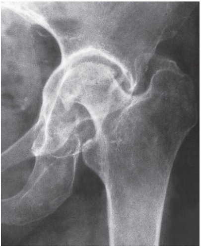
FIGURE 14.8 Rheumatoid arthritis. Anteroposterior radiograph of the left hip of a 59-year-old woman with advanced rheumatoid polyarthritis demonstrates the typical erosions of the femoral head and acetabulum. Note the lack of osteophytosis and the only very minimal reactive sclerosis.

Rice bodies. Bearing macroscopic similarity to grains of polished white rise, these small, usually uniform in size intraarticular or intrabursal loose bodies are commonly associated with rheumatoid arthritis, and are thought to represent a complication of chronic inflammatory process. Occasionally, they also may be seen in seronegative inflammatory arthritis, and even in tuberculous arthritis. These particles contain collagen, fibrinogen, fibrin, reticulin, elastin, mononuclear cells, blood cells, and some amorphous material. On radiography (Fig. 14.10), this condition occasionally can be mistaken for synovial chondromatosis (see Chapter 23). On MR T1-weighted images rice bodies exhibit intermediate signal intensity, whereas on T2 weighting they are only slightly hyperintense relative to muscle (Fig. 14.11).


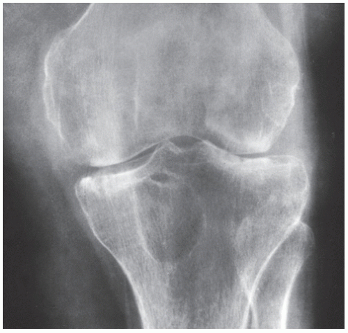
FIGURE 14.9 Rheumatoid cyst. Anteroposterior radiograph of the left knee of a 35-year-old woman with rheumatoid arthritis shows a large synovial cyst in the proximal tibia. Note also articular erosions and periarticular osteoporosis.


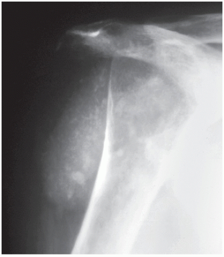
FIGURE 14.10 Rice bodies. Anteroposterior radiograph of the right shoulder of a 60-year-old woman with advanced rheumatoid arthritis demonstrates multiple rice bodies within subacromial-subdeltoid bursae complex.


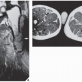
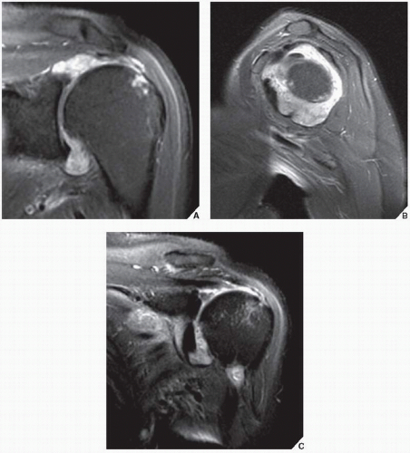
FIGURE 14.11 MRI of rice bodies. (A) Oblique coronal proton density-weighted, (B) sagittal proton density-weighted, and (C) oblique coronal T2-weighted fat-suppressed MR images of the left shoulder of a 66-year-old woman with rheumatoid arthritis show numerous rice bodies within the shoulder joint.


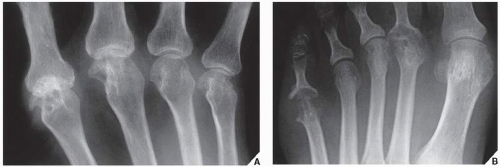
FIGURE 14.12 Rheumatoid arthritis of the small joints. Radiographs of the hand (A) and foot (B) of a 51-year-old woman with rheumatoid arthritis show typical erosions of the small joints.


Small Joint Involvement

Rheumatoid arthritis characteristically affects the small joints of the wrist, as well as the metacarpophalangeal and proximal interphalangeal joints of the hands and feet (Fig. 14.12). As a rule, the distal interphalangeal joints in the hand are spared, although in advanced stages of the disease even these may be affected. This latter point, however, is controversial, because some investigators believe that if the distal interphalangeal joints are involved, the condition may represent juvenile rheumatoid arthritis or another form of polyarthritis, not classic rheumatoid arthritis.

In addition to the characteristic changes exhibited in large joint involvement, the small joints may also show radiographic features specific for these sites.

Soft-Tissue Swelling. This earliest sign of rheumatoid arthritis usually has a fusiform, symmetric shape. It is periarticular in location and represents a combination of joint effusion, edema, and tenosynovitis.

Marginal Erosions. The earliest articular changes manifest as marginal erosions at so-called bare areas. These are the sites within the small joints that are not covered by articular cartilage. The most common locations for these erosions are the radial aspects of the second and third metacarpal heads and the radial and ulnar aspects of the bases of the proximal phalanges (Fig. 14.13). Synovial inflammation in the prestyloid recess, a diverticulum of the radiocarpal joint that is intimate with the styloid process of ulna, as Resnick pointed out, produces marginal erosion of the styloid tip.


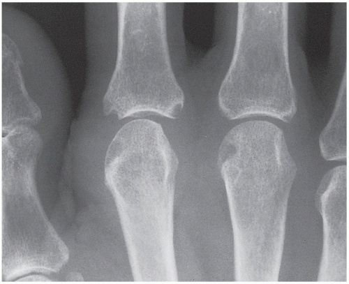
FIGURE 14.13 Rheumatoid arthritis. Typical erosions in the bare areas are seen in this 55-year-old woman with rheumatoid arthritis. Note also periarticular osteoporosis and soft-tissue swelling.

Joint Deformities. Although not pathognomonic for rheumatoid arthritis, certain deformations such as the swan-neck deformity and the boutonnière deformity are more often seen in this form of arthritis than in other inflammatory arthritides. The first of these represents hyperextension in the proximal interphalangeal joint and flexion in the distal interphalangeal joint, a configuration resembling a swan’s neck (Fig. 14.14). In the boutonnière deformity, the configuration is just the opposite, with flexion in the proximal joint and extension in the distal interphalangeal joint (Fig. 14.15). The word boutonnière is French for “buttonhole,” the term for this deformity deriving from the configuration of the finger while securing a flower to a lapel. A similar deformation of the thumb is called hitchhiker’s thumb.


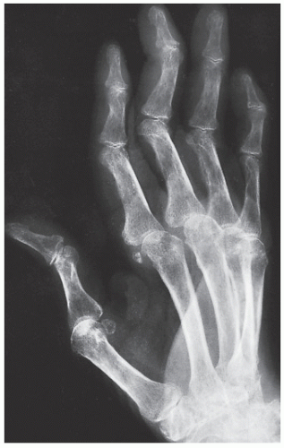
FIGURE 14.14 Rheumatoid arthritis. Oblique radiograph of the hand of a 59-year-old woman shows the swan-neck deformity of the second through fifth fingers. Note the flexion in the distal interphalangeal joints and the extension in the proximal interphalangeal joints, the hallmarks of this abnormality.


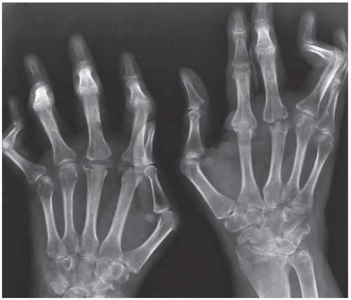
FIGURE 14.15 Rheumatoid arthritis. Dorsovolar radiograph of the hands of a 48-year-old woman with rheumatoid arthritis demonstrates the boutonnière deformity in the small and ring fingers of the right hand and in the ring finger of the left hand.

Moreover, subluxations and dislocations with malalignment of the fingers are common findings in advanced stages of rheumatoid arthritis. Particularly characteristic are ulnar deviation of the fingers in the metacarpophalangeal joints and radial deviation of the wrist in the radiocarpal articulation (Fig. 14.16). In far-advanced stages of rheumatoid arthritis, shortening of several phalanges may be encountered secondary to destructive changes in the joints associated with dislocations in the metacarpophalangeal joints. This deformity appears as a “telescoping” of the fingers, hence its name, main-en-lorgnette, from the French name for the telescoping type of opera glass (Fig. 14.17). An abnormally wide space between the lunate and the scaphoid may also be encountered in advanced stages of the disease secondary to erosion and rupture of the scapholunate ligament (Fig. 14.18); this phenomenon resembles the Terry-Thomas sign seen secondary to trauma
(see Fig. 7.82). Joint deformities are also often seen in the foot; the subtalar joint is frequently affected, and subluxation in the metatarsophalangeal joints often leads to deformities such as hallux valgus and hammertoes.


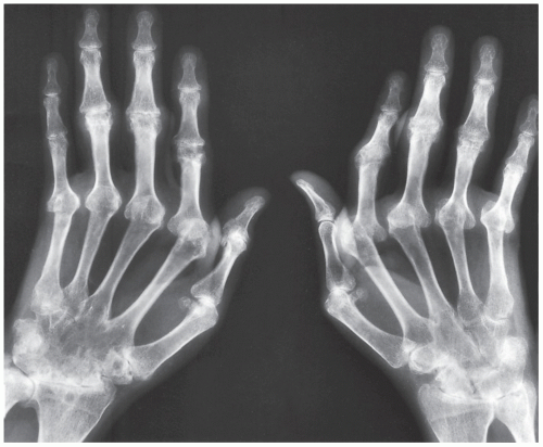
FIGURE 14.16 Rheumatoid arthritis. Dorsovolar projection of both hands of a 51-year-old woman shows subluxation in the metacarpophalangeal joints resulting in ulnar deviation of the fingers and radial deviation in the radiocarpal articulations. Note also ankylosis of the midcarpal articulations of the right hand.


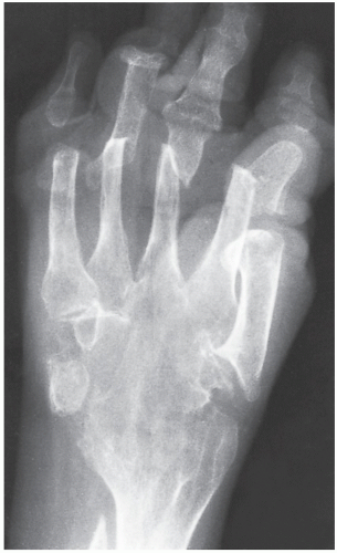
FIGURE 14.17 Rheumatoid arthritis. Dorsovolar view of the right hand of a 54-year-old woman with long-standing advanced rheumatoid arthritis demonstrates the main-en-lorgnette deformity. Note the telescoping of the fingers secondary to destructive joint changes and dislocations in the metacarpophalangeal joints. There is also ankylosis of the radiocarpal and intercarpal articulations and “penciling” of the distal ulna.

Joint Ankylosis. A rare finding that may be observed in advanced stages of rheumatoid arthritis is joint ankylosis, which is most commonly encountered in the midcarpal articulations (see Figs. 14.16 and 14.17). Ankylotic changes in the wrist are more common in patients with juvenile rheumatoid arthritis and with so-called seronegative rheumatoid arthritis.


Involvement of the Spine

The thoracic and lumbar segments are affected by rheumatoid arthritis only on rare occasions. The cervical spine, however, is involved in approximately 50% of individuals with this condition (Table 14.2). The most characteristic radiographic features of rheumatoid arthritis in the cervical spine can be observed in the odontoid process, the atlantoaxial joints, and the apophyseal joints. Erosive changes may be encountered in the odontoid process (see Fig. 12.36) and apophyseal joints (Fig. 14.19), whereas subluxation is a common finding in the atlantoaxial joint (see Fig. 12.37), frequently accompanied by vertical translocation of the odontoid process (also known as cranial settling or atlantoaxial impaction) (Figs. 14.20 and 14.21). The most frequent abnormality is laxity of the transverse ligament connecting the odontoid to the atlas. This laxity becomes apparent on the radiograph obtained in the lateral view of the flexed cervical spine, is expressed by subluxation in the atlantoaxial joint (Fig. 14.22), and is frequently accompanied by cephalad migration of the odontoid process. This complication often requires surgical intervention, and the most common procedure to correct this is posterior fusion.


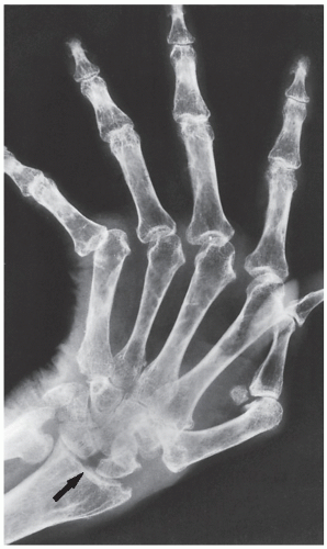
FIGURE 14.18 Rheumatoid arthritis. Dorsovolar view of the hand of a 60-year-old woman shows a gap between the scaphoid and lunate (arrow), indicating destruction of the scapholunate ligament. Note also the subluxation in the metacarpophalangeal joints resulting in ulnar deviation of the fingers.

Severe involvement of the apophyseal joints leads to subluxations. In extremely rare cases, in a manner similar to that in juvenile rheumatoid arthritis, the apophyseal joints may ankylose. The other structures occasionally affected by rheumatoid process are the intervertebral disks and adjacent vertebral bodies, which become involved as a result of synovitis extending from the joints of Luschka. Only a small percentage of patients with cervical disease may have cervical myelopathy. MRI is an ideal modality to evaluate spinal cord involvement in these patients (see Fig. 14.21).

Only gold members can continue reading. Log In or Register to continue

Stay updated, free articles. Join our Telegram channel

Jul 24, 2016 | Posted by in MUSCULOSKELETAL IMAGING | Comments Off on Inflammatory Arthritides

Full access? Get Clinical Tree

Get Clinical Tree app for offline access