9 Spleen
 Organ Boundaries
LEARNING GOALS
 Locate and identify the spleen.
 Demonstrate the spleen in its entirety.
Locating the spleen
The spleen lies posterolaterally below the left costal arch.
Barriers to scanning
The front of the spleen is completely covered by the stomach. Ribs, lung, and the left colic flexure are obstacles to scanning from the back and side (Fig. 9.1).
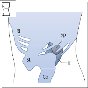
Fig. 9.1 Locating the spleen. The stomach (St), colic flexure (Co), and ribs (Ri) are barriers to scanning. Sp = spleen, K = kidney.
Optimizing the scanning conditions
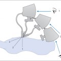
Given the difficulties in visualizing the spleen, the subject should be positioned on the right side with the left arm stretched up over the head (Fig. 9.2). Unlike the other upper abdominal organs, the spleen is usually easier to scan during expiration. Occasionally it is helpful to examine the spleen with the subject standing.
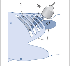
Fig. 9.2 Demonstrating the spleen. Note that the transducer is aligned parallel to the intercostal spaces, and the beam is directed cephalad. This will display, somewhat indistinctly, a triangular section of the spleen. Sp = spleen, K = kidney, Pl = pleura.
Organ identification
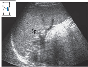
The spleen is located between the middle and posterior axillary line. Place the transducer in an intercostal space at that level and align it with the direction of the ribs (Fig. 9.3). As you can see from Figures 9.1 and 9.2, you must find an intercostal approach that is below the lung and then scan upward at a relatively steep angle.
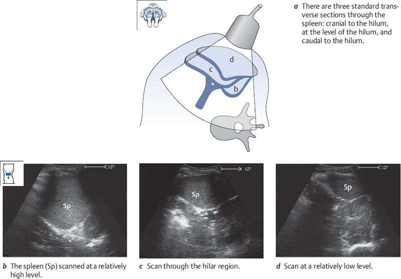
Imaging the entire spleen
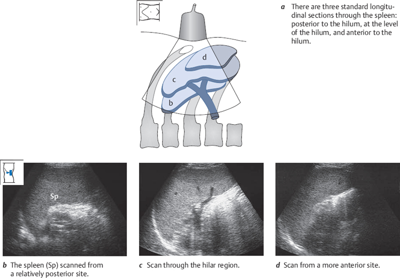
Preliminary note: we mainly use a systematic sequence of longitudinal and transverse sections in this book to define sonographic anatomy. We must deviate from this system somewhat for imaging the spleen, because the position of the ribs makes it difficult to perform standard longitudinal scans. So when we refer to longitudinal sections in this chapter, we mean that they are almost longitudinal (Fig. 9.4a).
Defining the spleen in longitudinal sections
Image the spleen in longitudinal section, and angle the transducer in an effort to obtain the best possible view. Now angle the scan posteriorly and see how the section of the spleen becomes smaller (Fig. 9.4b). Angle the transducer back toward the front, and watch the hilar vessels come into view (Fig. 9.4c). Continue scanning anteriorly until the section of the spleen becomes smaller again (Fig. 9.4d) and finally disappears.
Defining the spleen in transverse sections
Image the spleen in the familiar longitudinal section and rotate the transducer under vision to a transverse scan (Fig. 9.5a). Scan upward by angling the transducer cephalad, and then scan all the way down through the spleen in transverse sections (Fig. 9.5b–d).
 Organ Details
LEARNING GOALS
 Evaluate the shape of the spleen.
 Determine the size of the spleen.
 Evaluate the echo pattern of the spleen.
Shape of the spleen
In shape, the spleen resembles a spherical segment whose convexity is related to the diaphragm superiorly, posteriorly, and laterally. It has impressions for the kidney (posteromedially), stomach (anteromedially), and colon (inferiorly) (Fig. 9.6). The hilar vessels are located between the stomach and kidney (see Fig. 9.26a). The spleen is somewhat variable in its shape, however; it may be elongated or plump and occasionally shows deep constrictions (Fig. 9.7).
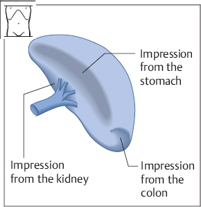
Fig. 9.6 Frontal view of the spleen. The surface of the spleen bears impressions for the stomach, colon, and left kidney.
Determining the size of the spleen
KEY POINT
The spleen normally measures 11–12 cm in length and 4 cm in depth.
For routine examinations, it is sufficient to measure the spleen in two dimensions. Define the spleen by scanning longitudinally from the flank as before. Angle the scan until it displays the splenic hilum. Measure the greatest longitudinal diameter in this view and the perpendicular diameter from the surface of the spleen to the hilum (Fig. 9.8).
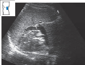
Fig. 9.8 Measuring the size of the spleen in a longitudinal flank scan. The normal dimensions are 11–12 cm by 4 cm.
Enlarged spleen
Related posts:
Stay updated, free articles. Join our Telegram channel

Full access? Get Clinical Tree


