Overview
Location
• Except for its medial hilum, the spleen is intraperitoneal (enclosed in the sac formed by the parietal peritoneum) in the posterolateral section of the left upper quadrant (or left hypochondrium), beneath the ninth to eleventh intercostal spaces.
• The spleen lies posterior and lateral to the tail of the pancreas, fundus and body of the stomach, and posterior to the splenic flexure (sharp bend between the transverse and descending colon in the left upper quadrant, anterior to the spleen).
• The medial portion of the spleen is superior and lateral to the left kidney.
• The spleen lies inferior and adjacent to the diaphragm, putting it in close proximity to the left pleural cavity.
Anatomy
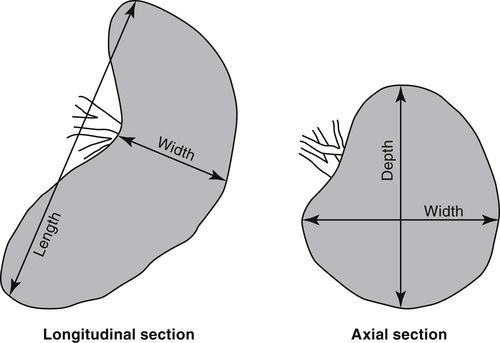
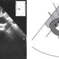
• The size of the spleen is variable, but is considered normal when it appears about the same size as the adjacent left kidney. Generally, it should be no longer than 12 or 13 cm and no deeper (anteroposteriorly) than 7 or 8 cm. The correct measurement caliper placement is demonstrated in Figure 10-1.
• The shape of the spleen is variable, but it is basically ovoid and may resemble a half moon or crescent. Thus, its superoposterior surface (adjacent to the diaphragm) is convex and smooth and its inferomedial surface (in contact with the stomach, left kidney, splenic flexure, pancreas tail) is concave and indented or nodulous. The splenic hilum (entry and exit route for the arterial, venous, and lymphatic vessels and nerves) is located within this concavity. Note that if the spleen becomes enlarged, it loses its concave appearance and becomes more round.
• The splenic vein exits the hilum and heads across the body toward the midline, running behind (posterior to) the tail and body of the pancreas to join the superior mesenteric vein at a point just posterior to the pancreas neck to form the portal vein. This location is commonly referred to as the portal-splenic confluence.
• The spleen receives blood from the splenic artery that branches from the celiac trunk of the abdominal aorta and runs left lateral and just superior to the body and tail of the pancreas, to enter the splenic hilum. In many cases, the artery may divide into two or three smaller branches before entering the spleen.
Physiology
• Although not essential to life, the spleen filters foreign material from the blood and forms antibodies.
• The spleen is a large mass of lymphatic tissue (reticular connective tissue) that is part of the reticuloendothelial system, an essential part of the immune system. It is composed of phagocytic cells capable of consuming substances, such as bacteria and viruses, making them incapable of causing harm to the body. They also engulf old cells and abnormal cells, clearing the body of their harmful presence.
• The spleen also breaks down hemoglobin, is a blood reservoir, and is important for hematopoiesis (red blood cell formation) in the fetus or when there is severe anemia (marked loss of the number of red blood cells produced in bone marrow; symptoms may include fatigue, shortness of breath, irregular heartbeat).
Sonographic Appearance
• The normal adult spleen appears homogeneous, with medium-level echoes and even texture that is described as isosonic or slightly hypoechoic when compared to the normal liver, and hyperechoic relative to kidney parenchyma. In some cases, small vascular branches can be seen interspersed within the spleen. They appear as anechoic, round or tubular structures. Arterial walls usually appear brighter than venous walls; however, the larger venous structures can clearly be distinguished from the smaller arterial branches at the level of the splenic hilum. Occasionally, small, bright reflections may be visualized throughout the spleen that represent calcified granulomatous inclusions or calcifications of small arterial walls.
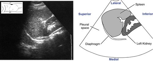
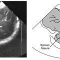
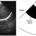
• The orientation of the spleen is vertically obliqued in the body, therefore longitudinal and long axis views are seen in oblique sagittal scanning planes and oblique coronal scanning planes as demonstrated in the image on the following page. This normal spleen appears crescent in shape and hyperechoic compared to the adjacent left kidney. Notice the spleen’s smooth outer convexity and its relationship to the diaphragm and pleural space superiorly.
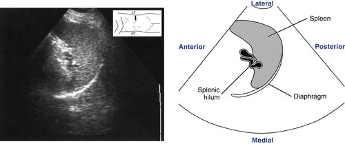
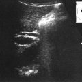
• Axial sections of the spleen are visualized in transverse scanning planes. The following is a transverse scanning plane image from a left lateral approach. It demonstrates an axial section of the spleen that includes the splenic hilum. Note the normal homogeneous appearance and even texture of the spleen.
Normal Variants
• Accessory spleen:
• Splenic tissue found separate from the organ, most often at the splenic hilum. Sonographic appearance is the same as normal splenic tissue.
• Asplenia:
• Rare absence of the spleen, often associated with congestive heart disease.
Preparation
Patient Prep
• None.
Transducer
• 5.0 MHz for intercostal or lateral subcostal scanning approaches.
• 3.0 or 3.5 MHz for anterior or posterior scanning approaches.
Breathing Technique
• Deep, held inspiration. Deep inspiration causes the spleen to descend, making it easier to visualize sonographically.
Patient Position
• Right lateral decubitus.
• Supine, sitting semierect to erect, and prone as needed.
Related posts:
Stay updated, free articles. Join our Telegram channel

Full access? Get Clinical Tree


