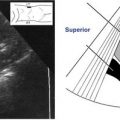
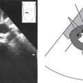
A, Illustrates the TV transducer position and sagittal plane field of view. B, Depicts the rotation of the image as seen on the display monitor. C, Illustrates a longitudinal section of the uterus in a TV sagittal plane. The apex of the image on the display monitor corresponds to the anatomy closest to the face of the transducer. In TV sonography, the near field and left side of the sagittal plane image generally correspond to the inferoposterior region of the true pelvis. The far field and right side of the sagittal plane image generally correspond to the anterosuperior region of the true pelvis. D, Longitudinal section of the uterus taken in a TV sagittal scanning plane. Note how the section of uterus fills the screen, limiting the overall view of the pelvis but providing increased anatomic detail of the uterus. (∗Denotes corresponding locations in A and C.)
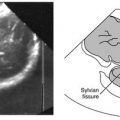
A, Illustrates the TV transducer position and coronal plane field of view. When the bladder is empty, the fundus of the typical anteverted (anteflexed) uterus tilts forward toward the anterior abdominal wall. Therefore, in TV imaging the uterus is seen in short axis from a coronal plane. B, Depicts the rotation of the image as seen on the display monitor. C, Illustrates an axial or short-axis section of the uterus in a TV coronal plane. The apex of the image on the display monitor corresponds to the anatomy closest to the face of the transducer. In TV sonography, the near field and left side of the coronal plane image generally correspond to the inferolateral region of the true pelvis. The far field and right side of the coronal plane image generally correspond to the superolateral region of the true pelvis. D, Axial or short-axis section of the fundus of the uterus taken in a TV coronal scanning plane. Note how the section of uterus fills the screen, limiting the overall view of the pelvis but providing increased anatomic detail of the uterus. (∗Denotes corresponding locations in A, B, and C.)
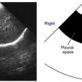
Most TV imaging is performed from the standard inferior approach as explained in Figures 12-1 and 12-2. Alternatively, manipulation of the TV transducer causes variation from the standard TV orientation. For example, these illustrations show that when the transducer is lifted anteriorly, toward the pubic symphysis, the sound beam is directed more posteriorly making the near and far fields of the TV sagittal plane image correspond to anterior and posterior regions of the pelvis instead of inferior and superior regions (from an inferior approach). The left and right sides of the image now correspond more closely to the superior and inferior regions of the pelvis instead of anterior and posterior (from an inferior approach). Utilizing a posterior TV approach also causes significant variation in image orientation. Consequently, image orientation for TV sonography can vary among institutions, authors, and ultrasound texts.
Transvaginal Female Pelvis Survey
Note
Because of the limited field of view, the uterus and adnexa are scanned in sections by slightly angling the inserted transducer in different directions.
Note
During the transvaginal evaluation, adnexal structures can be brought into view by using one hand to compress the lower abdominal wall while the other hand operates the transducer.
Uterus and Adnexa • Longitudinal Survey
Sagittal Plane • Inferior Approach
1. Begin scanning by slowly lowering the handle toward the floor to view a longitudinal section of the fundus of the uterus. Now move the transducer a little to the right, then to the left to evaluate its lateral margins. Note and evaluate the centrally located endometrial canal. If the bladder contains any urine, it will be seen anteriorly (on the left side of the imaging screen).
2. Withdraw the transducer slightly, and slowly lift the handle toward the ceiling to view the body and cervix of the uterus and the posterior cul-de-sac. Now move the transducer a little to the right, then to the left to evaluate the lateral margins. Note and evaluate the centrally located endometrial and endocervical canals.
3. After evaluating the uterus, continue the longitudinal survey to the adnexal regions. Very carefully reinsert the partially withdrawn transducer. Keep the transducer straight at the midline and lower the handle to relocate the uterine fundus and then the pelvic cavity region superior to the uterus. Now slowly move the transducer handle toward the patient’s left thigh in order to scan through the right adnexa. Return to midline; slowly move the transducer handle toward the patient’s right thigh to scan through the left adnexa.
Note
For a retroverted uterus, the uterine fundus is visualized by lifting the transducer handle towards the ceiling. It may also be helpful to rotate the probe 180 degrees and invert image orientation.
4. Repeat these lateral sweeps through the adnexal regions at the levels of the uterine body and cervix.
Uterus and Adnexa • Axial Survey
Coronal Plane • Inferior Approach
1. Following the longitudinal survey from the sagittal plane, rotate the transducer 90 degrees counterclockwise into the coronal plane.
2. Begin scanning by slowly lowering the handle of the inserted transducer toward the floor to evaluate the uterine fundus.
3. Withdraw the transducer slightly and slowly lift the transducer handle toward the ceiling to scan through the uterine body, cervix, and posterior cul-de-sac.
4. After evaluating the uterus, continue the axial survey through the adnexa. Moving the transducer handle toward the floor, relocate the uterine fundus. Now slowly move the transducer handle toward the patient’s left thigh to visualize the right adnexa, and very slowly move the transducer handle toward the ceiling in order to sweep through the area.
5. Return the transducer to the midline, then slowly move the handle toward the patient’s right thigh to visualize the left adnexa. By very slowly moving the transducer handle up and down, sweep through the area.
Right Ovary • Axial Survey
Coronal Plane • Inferior Approach
1. The ovaries are most easily evaluated by beginning in the coronal scanning plane. From initial insertion of the transducer in the sagittal plane, the transducer handle should be rotated 90 degrees counterclockwise into the coronal plane.
2. Begin scanning by putting the transducer in a right oblique position. This is done by slowly moving the transducer handle toward the patient’s left thigh, which angles the beam toward the right adnexa. Find the ovary by slightly moving the transducer handle up and down. Identify the adjacent iliac vessels.
3. When the ovary is located, very slightly move the transducer handle as far up and down as necessary to scan through the ovarian margins.
Right Ovary • Longitudinal Survey
Sagittal Plane • Inferior Approach
1. Still viewing the ovary in the coronal plane, slowly rotate the transducer 90 degrees clockwise to visualize the ovary in the sagittal plane.
Only gold members can continue reading. Log In or Register to continue