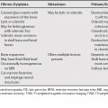
90 The differential diagnosis of splenic lesions is broad, but can be more easily approached if divided into solitary and multiple lesions. The next consideration is whether the lesion(s) are cystic, solid and nonvascular, or vascular. The vascular lesions can be distinguished from the solid nonvascular lesions by a greater degree of enhancement. Lymphangiomas are more common in children. They usually appear as single or multiple cystic lesions, sometimes multiloculated, which are often subcapsular in location. Curvilinear peripheral mural calcifications can be seen. Lymphangiomas are usually hypointense on T1-weighted magnetic resonance images (T1WI), but may be hyperintense from hemorrhage or proteinaceous material. They do not enhance.1 Daughter cysts can be seen either peripherally or throughout the lesion, resulting in a multilocular appearance. These daughter cysts can appear less dense on computed tomography (CT) and more hypointense on T1WI relative to fluid in the parent cyst. Other possible differentiating features include collapsed parasitic membranes within the cyst, a low-intensity rim around the cyst secondary to a fibrous capsule, and cyst wall calcification.2 Splenic metastases, which are necrotic or mucinous, can appear cystic. Serosal implants from ovarian cancer can appear as peripheral cystic lesions. Hemangiomas are the most common benign primary neoplasm of the spleen, and the second most common focal lesion (after cysts). They are most often seen in patients between the ages of 30 and 50. There may be a slight male predilection. Splenic hemangiomas have been associated with Klippel-Trenaunay-Weber syndrome (the presence of phleboliths in or adjacent to the spleen suggests a visceral angiomatosis syndrome).3 Most hemangiomas are smaller than 2 cm. There is occasional calcification. Hemangiomas are usually homogeneously hyperintense on T2-weighted magnetic resonance images (T2WI; more homogeneous than hamartomas). Hemangiomas have a variable enhancement pattern. They can have a similar enhancement pattern to liver hemangiomas (progressive centripetal enhancement with persistent delayed enhancement), but differ from liver hemangiomas in that most do not have peripheral enhancing nodules. Some hemangiomas demonstrate mottled enhancement. Smaller hemangiomas may enhance homogeneously.1,4 Splenic hamartomas are rare lesions composed of malformed red pulp elements. They can be seen in any age and have no sex predilection. They are associated with tuberous sclerosis. Hamartomas usually have well-defined margins and occasionally contain calcification and fatty components. They are usually heterogeneously hyperintense on T2WI (but sometimes can be hypointense on T2WI if fibrous). The enhancement pattern of hamartomas can be helpful in differentiating hamartomas from other lesions. Hamartomas often demonstrate diffuse heterogeneous early contrast enhancement and homogeneous prolonged enhancement.1,5 Compared with hemangiomas, hamartomas have greater heterogeneity on T2WI and more heterogeneous4
Splenic Lesions
Cystic Splenic Lesions
Lymphangioma
Echinococcal Cyst
Cystic Metastasis
Vascular Splenic Lesions
Hemangioma
Hamartoma
![]()
Stay updated, free articles. Join our Telegram channel

Full access? Get Clinical Tree


