8 Stomach, Duodenum, and Diaphragm
TIP
The liver provides an acoustic window for scanning the stomach and duodenum.
LEARNING GOALS
 Identify the stomach and duodenum.
 Identify the area of the diaphragm pierced by the aorta and vena cava.
The stomach and duodenum are usually considered barriers to scanning and are not always specifically identified and examined as objects of interest. While it is true that the stomach and duodenum are not classic ultrasound organs, often they are not too difficult to examine, even without special patient preparations, if you know where to look. Of course, gastrointestinal scanning is basically a task for the advanced sonographer. However, we include the stomach and bowel in this introduction to emphasize that they are not merely obstacles but are part of the upper abdominal anatomy that is accessible to deliberate examination. At the same time, we would advise beginners to be very cautious in interpreting their findings.
A familiarity with the diaphragm in the area where it is pierced by the aorta and vena cava is of some importance, since the diaphragm in that area may be confused with the right adrenal gland or may be misinterpreted as a vascular structure.
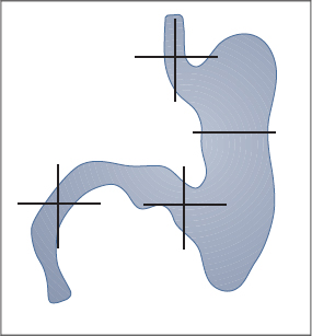
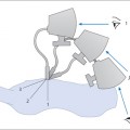
Figure 8.1 shows an anterior view of the stomach and duodenum as they appear in anatomical textbooks.
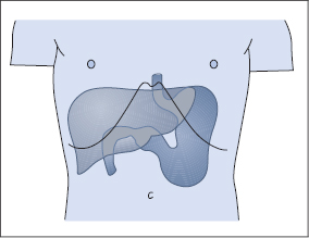
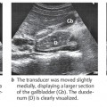
The structures that often can be seen clearly with ultrasound are the cardia and gastroesophageal junction, the antrum, and the first and second parts of the duodenum (Figs. 8.2, 8.3). The liver provides an acoustic window for scanning these structures. It is far more difficult to obtain a clear view of the fundus and body of the stomach by scanning from the front of the abdomen or through the spleen.
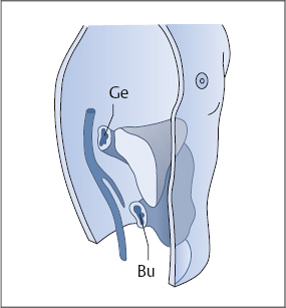
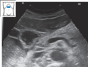
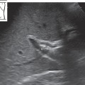
Fig. 8.2 Oblique longitudinal scan. This plane demonstrates the gastroesophageal junction (Ge) and the junction of the stomach and duodenal bulb (Bu).
 Organ Details
Stomach wall
If the scanner has good resolution and conditions are favorable, five layers can be distinguished in the stomach wall (Fig. 8.4):
 The echogenic interface between the lumen and mucosa
 The hypoechoic muscularis mucosae
 The echogenic submucosa
 The hypoechoic muscularis externa
 The echogenic outer surface of the serosa
These five layers cannot always be clearly identified, however. The best section for this purpose is one through the antrum. Often only three layers can be recognized: the echogenic inner and outer layers and the hypoechoic middle layer (as shown in Fig. 8.21a).
Changes in the stomach wall
It would be beyond our scope to explore the ultrasound diagnosis of benign and malignant changes in the stomach wall. Figure 8.5 shows an example of a stomach wall lesion that can be identified with ultrasound.
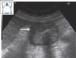
Abb. 8.5a Gastric carcinoma. Asymmetrical, hypoechoic expansion of the stomach wall in the antral region (→).
 Organ Boundaries and Relationships
Esophagus and cardia
Defining the gastroesophageal junction in longitudinal section
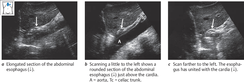
Generally the cardia is best demonstrated in an upper abdominal longitudinal scan that displays the stomach between the liver and the aorta. Center the transducer very high in the epigastrium (Fig. 8.6b). Visualize the aorta. Now angle the transducer to scan longitudinally into the upper abdomen. Tilt it slightly to the right, and you will obtain an elongated section of the esophagus (Fig. 8.6a). Now angle the transducer to the left, and you will see the esophagus merge with the gastric cardia (Fig. 8.6c).
Relationships of the esophagus and cardia
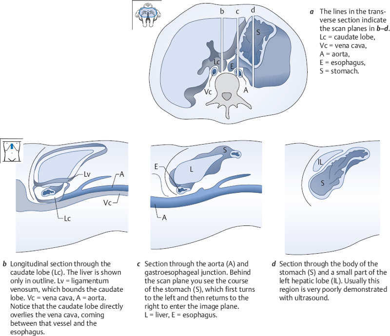
Figure 8.7 illustrates the structures that surround the gastroesophageal junction.
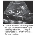
Scan longitudinally over the aorta and display the familiar section (Fig. 8.8a
Related posts:
Stay updated, free articles. Join our Telegram channel

Full access? Get Clinical Tree


