Chapter 110
Subglottic Hemangioma
Epidemiology
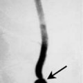
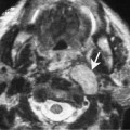
Hemangiomas may occur anywhere in the larynx but have a predilection for the subglottic region. They are slow-growing benign lesions and typically present within the first year of life, with 85% of masses presenting before 6 months of age. Subglottic hemangiomas (SHs) are more common in females with a female to male ratio of 2:1. They are more common on the left; however, they may occur on the right or be bilateral. Cutaneous hemangiomas have been reported in association with SH in 50% of cases.
Clinical Findings
Patients with SH typically present with signs or airway obstruction with inspiratory or biphasic stridor. Other symptoms include dyspnea, cyanotic episodes, hoarseness, cough, and difficulty feeding. Continued growth may cause significant airway obstruction. The symptoms are often exacerbated by excitement, crying, or respiratory tract infection. Occasionally, symptoms may progress to acute respiratory distress. The typical endoscopic appearance is that of a red or blue submucosal mass situated in the subglottis. The mass is compressible and may extend into the posterior commissure or upper trachea.
Pathology