20 Superior Mesenteric Artery and Colic Arteries
K.I. Ringe
The gastroduodenal arteries (see Chapter 14 and 17), the jejunal, and ileal arteries are not depicted, but the arteries supplying the colon, cecum, and appendix are outlined in detail.1–16 The ileocolic artery is the most variable of the colic branches from the superior mesenteric artery. Accessory middle colic arteries are of clinical importance, since in such cases the arterial blood supply of the colon by the superior mesenteric artery is extended to the left colic flexure or even as far as the descending colon. Accessory middle colic arteries have been described as branches of the gastroduodenal or left gastroepiploic arteries. if accessory branches from the inferior mesenteric artery are present, arteries supply the transverse colon up to the right colic flexure. Normally the anastomosis along the colon formed by different arches (marginal artery) is large enough to ensure a sufficient collateral circulation (anastomosis of Riolan). This anastomosis between the superior and inferior mesenteric arteries is absent in approximately 2% of all cases, and in a further 3% it is very small. The arch between the right colic and the ileocolic arteries seems to be absent in approximately 6% of all cases.1,2,9,13,17–30
20.1 Three Colic Arteries Branch from the Superior Mesenteric Artery (67%)

Fig. 20.1 The ileocolic, right, and middle colic arteries are independent branches of the superior mesentery artery (24%). Schematic (a), DSA of the superior mesenteric artery (b), and coronal MIP CT of the superior mesenteric artery (c). 1 Right colic artery; 2 middle colic artery; 3 superior mesenteric artery; 4 ileocolic artery.

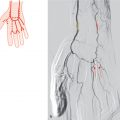
Fig. 20.2 The ileocolic and right colic arteries form a trunk (20%). Schematic (a) and DSA of the superior mesenteric artery of two patients (b,c). 1 Right colic artery; 2 middle colic artery; 3 superior mesenteric artery; 4 ileocolic artery.

Fig. 20.3 The right and middle colic arteries from a common trunk (22%). Schematic (a) and DSA of the superior mesenteric artery (b). 1 Right colic artery; 2 middle colic artery; 3 superior mesenteric artery; 4 ileocolic artery.

Fig. 20.4 The ileocolic, right, and middle colic arteries form a common trunk (1%). Schematic.
20.2 Two Colic Arteries Branch from the Superior Mesenteric Artery (15%)

Fig. 20.5 The right colic artery is absent (10%). The ileocolic and middle colic arteries in such cases normally branch separately from the superior mesenteric artery. Schematic (a) and DSA of the superior mesenteric artery of two patients (b,c). 1 Ileocolic artery; 2 middle colic artery; 3 superior mesenteric artery.

Fig. 20.6 The middle colic artery is absent (5%). The ileocolic and right colic arteries branch separately in three-fourth of all cases; otherwise they form a common trunk. Schematic (a) and DSA of the superior mesenteric artery (b). 1 Right colic artery; 2 superior mesenteric artery; 3 ileocolic artery.
20.3 Accessory Colic Arteries (18%)

Fig. 20.7 Two right colic arteries normally from common trunks with the middle colic and ileocolic artery (6%). Schematic (a) and DSA of the superior mesenteric artery (b). In addition, the patient has a replaced right hepatic artery from the superior mesenteric artery. 1 Right hepatic artery; 2 superior mesenteric artery; 3 ileocolic artery; 4 right colic artery 1; 5 right colic artery 2; 6 middle colic artery.
Related posts:


Stay updated, free articles. Join our Telegram channel

Full access? Get Clinical Tree


