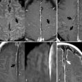
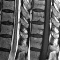
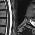
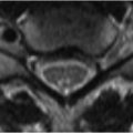
1 Supratentorial Brain Neoplasms Astrocytomas are the most common primary intraaxial tumor, arising predominantly supratentorially in adults and infratentorially in children. The World Health Organization’s (WHO) criteria divide astrocytomas into four grades: grade 1 is circumscribed astrocytoma (usually pilocytic astrocytomas); grade 2 is low-grade astrocytoma; grade 3 is anaplastic astrocytoma; and grade 4 is glioblastoma multiforme (GBM). The magnetic resonance imaging (MRI) appearance of grade I astrocytomas is typified by increased signal intensity (SI) on T2-weighted images (T2WIs) and decreased signal intensity on T1WI, reflecting increased extracellular fluid due to abnormal capillary walls. These lesions typically do enhance. Unlike other astrocytomas, the prognosis for grade 1 lesions is usually favorable, and they are often cured by resection alone. Juvenile pilocytic astrocytomas (JPAs), the most common grade 1 lesion, will be discussed in Chapter 2. Grade 2 astrocytomas demonstrate a relatively homogeneous appearance with well-defined borders on MRI (Fig. 1.1A,B)–an appearance that may belie an infiltrative pathology and poor prognosis. These tumors may grow large enough to exhibit significant mass effect, such as midline shift (mild left-to-right in the instance of Figs. 1.1A,B), but they generally lack the degree of edema (Fig. 1.1A) seen in higher-grade counterparts. Their lack of enhancement also helps distinguish them from a GBM. MR perfusion scans may help distinguish borderline cases, with higher-grade tumors demonstrating elevated cerebral blood volume. Grade 2 tumors may arise from multiple cell lines. The mass in Figs. 1.1A,B
![]()
Stay updated, free articles. Join our Telegram channel

Full access? Get Clinical Tree


