The adult human brain has a highly complex external morphology, and this is particularly true of the cerebral hemispheres. The clinical neuroimager needs to know the normal patterns of cortical gyri and their associated sulci in order to make accurate anatomic diagnoses that will assist in functional assessment and/or surgical planning. Someone looking at the surface of the adult brain for the first time likely would be convinced by the apparent randomness of the convoluted surface. However, it becomes apparent that the gyri/sulci form patterns that are common among individuals and, although variations exist, a large number of recurring themes can be found. It is important for anyone trying to understand the development of fetal cerebral hemispheres for diagnostic purposes to have a deep understanding of the final adult patterns and common variations. It is also necessary to appreciate the gestalt of being able to understand the surface anatomy of the brain and applying that knowledge when interpreting cross-sectional imaging studies. Naidich et al. have provided many illuminating publications on the subject, and the interested reader is directed to their work.
Before 16 weeks’ gestational age the fetal human cerebral hemispheres are effectively smooth and featureless. In contrast, the overall degree of sulcation at birth is effectively the same as the adult pattern. The huge changes in the external morphology of the brain that occur between those two time points are due to the development of the cerebral cortex and the massive numbers of neurons and glia that migrate there from the germinal matrices. The gyral convolutions produce a greater surface area per unit volume compared with the smooth, agyric cortex present in many other mammals. Indeed, the gyric human cerebral cortex is estimated to have three times the surface area as an agyric brain of the same volume. The major sulci of the brain tend to appear in an ordered and predictable sequence, and the person interpreting fetal magnetic resonance (MR) images should be aware of the normal patterns and schedules of appearance. However, the patterns are only approximations, and one should not expect to be able to define with any degree of accuracy the gestational age of a fetus based on the sulcal patterns. Biologic variation is one issue, and the mechanisms for estimating the dates of a pregnancy have wide margins of error. In addition, the possible significant differences in the degree of sulcation between the two hemispheres within the same individual are well documented.
The purpose of this section is to show the development of the surface cortical patterns of the fetal brain between 19 weeks’ gestational age and term. We recommend that you refer back to this section when studying the cross-sectional images of the appropriate gestational age in Section 2 or the neonatal cases in Section 3 because an understanding of sulcation both on cross-sectional imaging and on representations of surfaces is necessary. This section begins with a discussion of the appearances of the major cortical sulci that may be described as mature” or adult pattern. This section uses the surface projections of the developing fetal brain from the Larroche atlas.
Anatomy of the Sulci and Fissures in the “Mature” Supratentorial Brain
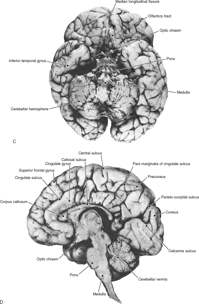
The cerebral hemispheres are separated from each other in the midline by the median (great) longitudinal fissure and its contents: the pia and arachnoid mater with the intervening subarachnoid space that overlie both cerebral hemispheres, and two layers of dura mater that are fused for the most part as the falx cerebri. The inferior sagittal sinus is contained within the free inferior border of the falx, whereas superiorly the two leaves of dura separate to contain the superior sagittal sinus ( Figure 1-1 ). The falx is attached to the crista galli anteriorly, where it is quite narrow, but it widens as it sweeps posteriorly and eventually attaches along the midline of the tentorium cerebelli. The drainage of venous blood in the sagittal sinuses normally is from anterior to posterior; therefore the structure increases in size passing posteriorly to accommodate for increasing drainage from the cortical veins. These features are well shown on coronal MR imaging.

The surfaces of the cerebral hemispheres show many convolutions consisting of cortical gyri separated by sulci of varying sizes. Most of the sulci are prominent and easily delineated in whole brain preparations, and many of them are constant between individuals. The cerebral cortex and associated white matter form four lobes in each hemisphere (frontal, temporal, parietal, occipital), and those lobes are (incompletely) defined by prominent, relatively constant sulci. The appearances of sulci on imaging studies can be appreciated only if the anatomy of the meninges is understood. The innermost layer of the meninges, the pia mater, is closely adherent to the surface of the brain at all sites. In contrast, the thicker arachnoid mater encompasses the brain without extending into the recesses. The subarachnoid space lies between the two, contains cerebrospinal fluid (CSF), and usually is quite thin. However, some regions contain local dilatations of the subarachnoid space with large pools of CSF. One such region is the basal cisterns related to the inferior surface of the brain; another is the space between adjacent cortical gyri. Thus the cortical sulci have the same intensity as the fluid within the ventricles on all sequences (e.g., high signal on T2-weighted images), and their shape is dependent solely on the shape of the adjacent gyri. The major sulci and associated brain structures of a fetus of 40 weeks gestational age are shown in Figure 1-2 .

Major Sulci Responsible for Defining Lobar Anatomy
These consist of the lateral (sylvian) sulcus, central sulcus, and parieto-occipital sulcus. For the most part the lobar anatomy is best defined on the lateral surface of the brain by the lateral and central sulci.
Lateral Sulcus
The lateral sulcus is a deep fissure that is first identified on the inferior surface of the brain close to the anterior perforated substance but becomes most visible on the lateral surface where it separates the frontal and parietal lobes from the temporal lobe. The frontal lobe is separated completely from the temporal lobe, whereas the posterior aspects of the parietal and temporal lobes remain in continuity without a well-defined external border. The parts of the frontal, temporal, and parietal lobes that protrude into and surround the lateral fissure are called the opercula. The anatomy of the lateral sulcus on the lateral surface of the brain is complicated as it divides into three rami: anterior horizontal, anterior ascending, and posterior. These can be seen well on MR imaging that allows nonorthogonal plane reformation of volume data ( Figure 1-3 ). The anterior horizontal ramus protrudes into the inferior frontal gyrus running horizontally and anteriorly. The anterior ascending ramus runs vertically into the same gyrus and defines the pars triangularis portion of the inferior frontal gyrus anterior to the ascending ramus and the pars opercularis posteriorly. The posterior ramus extends posteriorly and slightly superiorly for approximately 8 cm before dividing into the posterior ascending and posterior descending rami. Naidich et al. have shown that the anatomy of the subcentral gyrus is well seen on MR, and this topic is discussed in the section on locating the central sulcus.


The insula is defined as the cortical surface in the depth of the lateral fissure and is considered to be the “fifth cortical lobe” by some researchers. The mature insula has a complicated surface structure, which is best appreciated on whole brain preparations when the opercula have been removed (similar “virtual” procedures can be performed on T1-weighted volume data; Figure 1-4 ). The insula is pyramidal in shape, with its apex directed inferiorly and anteriorly. The apex is the only portion of the insula that is not bounded by the circular gyrus. The large central insular sulcus runs from the apex, superiorly and posteriorly to form larger anterior and smaller posterior surfaces. The posterior region usually is divided by a single sulcus to form two “gyri longi,” whereas the anterior area is inconsistently divided into three or four “gyri brevi.”

Central Sulcus
This prominent sulcus on the lateral aspect of the cerebral hemisphere barely extends onto the medial surface, if at all. The central sulcus separates the frontal and parietal lobes, and the frontal lobe can be completely delineated by the lateral and central sulci on the lateral surface of the brain. It takes a curved course posteriorly at approximately 70° towards the lateral sulcus but does not contact it. The postcentral sulcus lies approximately 1.5 cm posterior to the central sulcus and runs parallel to it. The correct localization of the central sulcus is hugely important on cross-sectional imaging as it defines the primary motor cortex anteriorly and the primary sensorimotor cortex posteriorly. This can be difficult and is best achieved on axial imaging as described in the section on the cingulate sulcus.
Parieto-occipital Sulcus
This is predominantly a feature of the posterior portion of the medial hemispheric surface, although it can extend onto the lateral surface for a short way in some cases. It runs inferiorly and slightly anteriorly, separating the precuneus of the parietal lobe and the cuneus of the occipital lobe before joining the calcarine fissure.
Note that a temporo-occipital sulcus exists on the inferior surface of the brain but has highly variable appearances.
Other Sulci of Importance for Fetal Imaging
Superior and Inferior Frontal Sulci
The lateral surface of the frontal lobe is indented by two sulci running in a broadly horizontal fashion, the superior and inferior frontal sulci. These demarcate the superior frontal gyrus (above the superior frontal sulcus), inferior frontal gyrus (below the inferior frontal sulcus), and middle frontal gyrus between the two. These are well shown on coronal MR images. The precise pattern of sulcation varies a great deal, but most frequently the superior frontal sulcus is deficient posteriorly, allowing continuity between the posterior parts of the superior and middle frontal gyri.
Cingulate Sulcus
The most prominent feature on the medial aspect of the anterior cerebral hemisphere is the cingulate sulcus. The majority of this sulcus is related to the frontal lobe, commencing below the rostrum of the corpus callosum and curving anteriorly and then posteriorly roughly parallel to the corpus callosum and delineating the cingulate gyrus. At a point approximately above the splenium of the corpus callosum, the cingulate sulcus curves upward into the parietal lobe to become the pars marginalis of the cingulate gyrus, which extends onto the superior portion of the lateral aspect of the hemisphere. As described by Naidich et al., this is a useful landmark for locating the central sulcus on cross-sectional imaging.
Superior and Inferior Temporal Sulci
The lateral aspects of the temporal lobes are subdivided in a fashion similar to the frontal lobes. Two horizontally directed sulci, the superior and inferior temporal sulci, divide the surface into three gyri, the superior, middle, and inferior temporal gyri. Posteriorly there exists an indistinct boundary between the temporal gyri and the parietal and occipital lobes.
Calcarine Sulcus
The calcarine sulcus is a feature of the medial surface of the occipital lobe. It is important because the visual cortex lies above and below the calcarine sulcus. It commences at the occipital pole and runs anteriorly to meet the parieto-occipital sulcus.
Collateral Sulcus
The collateral sulcus starts at the occipital pole on the inferior surface of the brain and runs anteriorly parallel to the calcarine sulcus. At its anterior extent it separates the parahippocampal gyrus from the more lateral portions of the temporal lobe. It may join the rhinal sulcus, but more often it remains isolated.
Location of the Central Sulcus on Cross-Sectional Imaging
There are many situations in clinical practice when it is necessary to demonstrate the central sulcus on cross-sectional imaging in order to locate the precentral and postcentral gyri with confidence. This can be done only with an understanding of the anatomy of other sulcal structures. This is best performed on axial imaging for the superior portion of the central sulcus and on parasagittal images for the inferior portion. The anatomy of cortical sulci is best appreciated in older adults in whom some volume loss of the brain results in prominence of the sulci. Conversely, the brains of most children have a relative paucity of CSF-containing structures on the surface, which can make appreciation of sulcal anatomy difficult. Neonates, however, often have prominent sulci (and ventricles), which assists with location of the following structures.
Superior Portion of the Central Sulcus
The key to locating the superior portion of the central sulcus is being able to find the pars marginalis portion of the cingulate sulcus and the postcentral sulcus. As previously described the main stem of the cingulate sulcus is best shown on sagittal imaging just off the midline. The anterior portion of the cingulate sulcus is directly superior to the cingulate gyrus and runs parallel to the corpus callosum. Above the posterior part of the body of the corpus callosum a branch of the cingulate sulcus arcs superiorly. This is the pars marginalis, and it extends onto the superior surface of the cerebral hemispheres for a short distance. As a result, the pars marginalis has a highly characteristic appearance on the superior sections of axial brain images. It appears as an anteriorly curved sulcus that, when both sides are viewed together, has been likened to Salvador Dali’s moustache. The position of the pars marginalis in relation to the center of the image is dependent on the angulation of the axial sections. For example, axial MR images usually are set parallel to the anterior/posterior commissural line, which broadly approximates to the plane of the anterior cranial fossa. In this situation the pars marginalis is situated close to the posterior edge of the superior-most axial images ( Figure 1-5 ). If a steeper angulation is made, as for X-ray CT of the brain (in order to reduce radiation dose to the lens of the eyes), the pars marginalis is much closer to the center of the field of view.