Postoperative Evaluation of Congenital Heart Disease


  • Cardiac magnetic resonance (CMR) can be used to detect and quantify the most common complications after repair of tetralogy of Fallot. CMR can accurately measure pulmonary regurgitant fraction (phase-contrast CMR) and assess right ventricular function (cine CMR) and morphology in these patients.


  • CMR can assess hemodynamic significance of recurrent or residual coarctation of the aorta.


  • After surgical correction for transposition of great arteries, CMR can be used to evaluate for possible complications of atrial switch and arterial switch procedures.


  • CMR can identify and estimated the severity of conduit stenosis and regurgitation after the Rastelli operation.


  • CMR can be used to assess for the most common complications associated with the Fontan procedure, which are conduit stenosis or thrombosis and dysfunction of the single functioning ventricle.


KEY POINTS


Case 1


A 28-year-old man presents with a recent history of shortness of breath and palpitation during exercise. Clinical examination is unremarkable, except for a diastolic cardiac murmur. Past medical history is significant for surgical correction of tetralogy of Fallot at the age of 4.


Pulmonary Regurgitation after Correction of Tetralogy of Fallot


Comments


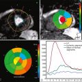
Pulmonary regurgitation is the most common complication observed after correction of tetralogy of Fallot, usually presenting clinically as intolerance to exercise. It is nearly always present in patients who underwent a transannular patch repair for correction of the right ventricular outflow tract stenosis and is often associated with dyskinesis or aneurysm on the patch area. Chronic pulmonary regurgitation eventually causes right ventricular dilatation and dysfunction. The CMR study can accurately measure pulmonary regurgitant fraction (phase-contrast CMR) and assess right ventricular function (cine CMR) and morphology in these patients ( Figure 16-1 ).


Figure 16-1


A, Double-inversion T1-weighted spin-echo MR image in the short-axis plane demonstrates the right ventricular outflow tract denuded of normal myocardium ( arrowheads ), consistent with status post a transannular patch repair for correction of tetralogy of Fallot. RV, right ventricle; LV, left ventricle. B-C, Short-axis steady-state free-precession (SSFP) cine images at end-diastole ( B ) and end-systole ( C ) show dyskinesis of the right ventricular outflow tract ( arrows ), a common complication after transannular patch repair. RV, right ventricle; LV, left ventricle. D, Inversion-recovery turbo spin-echo image in the short-axis plane obtained 10 minutes after the administration of gadolinium chelate. Note the area of delayed contrast enhancement in the right ventricular outflow tract (arrows), corresponding to the area of dyskinesis. The enhancement indicates the presence of fibrosis at the site of the surgical patch and/or adjacent nonviable myocardium. RV, right ventricle; LV, left ventricle. E, Velocity-encoded cine images obtained to quantify pulmonary regurgitant fraction in a plane perpendicular to the direction of blood flow in the main pulmonary artery. Oblique axial magnitude image (left), and phase images in systole (middle) and diastole (right) are shown. Phase contrast images show forward flow in systole ( dark voxels ) and retrograde flow in diastole ( bright voxels ) ( arrow ). F, Flow versus time curve displays the forward and retrograde flow in the pulmonary artery. Area under negative component of the curve yields a direct quantification of the volume of regurgitation. Pulmonary regurgitant fraction was calculated as 40%.


Case 2


A 30-year-old woman presents with intolerance to exercise and shortness of breath. Clinical examination is noticeable for both systolic and diastolic murmurs. Patient underwent correction of tetralogy of Fallot with infundibulectomy and pulmonary valvotomy at the age of 2.


Residual Pulmonary Stenosis after Correction of Tetralogy of Fallot


Comments


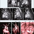
A common complication after correction of tetralogy of Fallot with infundibulectomy is residual infundibular and/or valvular pulmonary stenosis, which is usually associated with a mild to moderate degree of pulmonary regurgitation. Branch pulmonary artery stenosis is a common coabnormality seen in patients with tetralogy of Fallot and is often diagnosed later in life, after the initial surgical correction has been performed. A comprehensive CMR study allows for evaluation of morphology of stenosis and quantification of stenotic pressure gradient and ventricular function ( Figure 16-2 ).


Figure 16-2


A, Double-inversion T1-weighted spin-echo MR image in the sagittal plane demonstrates residual valvular pulmonic stenosis ( arrowhead ), a common complication after infundibulectomy and valvotomy to correct tetralogy of Fallot. Note the thick right ventricular myocardium and trabeculations ( asterisk ) consistent with chronic pressure overload. RV, right ventricle; LV, left ventricle. B-C, SSFP cine images in the right ventricular outflow tract plane were obtained during systole ( B ) and diastole ( C ). A flow void is identified distal to the pulmonary valve in systole, indicating a high velocity jet caused by pulmonary stenosis ( arrow ). During diastole, a high velocity jet is seen below the level of the pulmonary valve ( arrow ), consistent with associated pulmonary regurgitation. RV, right ventricle; LV, left ventricle. D-E, VEC-MR images were obtained parallel to the direction of blood flow in the main pulmonary artery. Magnitude (d) and phase (e) images are shown. A region of interest was placed in the phase image to interrogate the peak velocity distal to the pulmonary valvular stenosis, which was measured as 5 m/s. Using the modified Bernoulli equation (ΔP = 4 × PV 2 , where P = pressure in mm Hg and PV = peak velocity in m/s) a pressure gradient of 100 mm Hg was estimated across the stenosis. RV, right ventricle; LV, left ventricle; PA, pulmonary artery. F-G, Double-inversion T1-weighted spin-echo MR image in the axial plane ( F ) shows a long segment stenosis of the right pulmonary artery (arrow). VEC-MR images were obtained perpendicular to the direction of blood flow in the branch pulmonary arteries (white lines) for calculation of differential pulmonary blood flow. Flow versus time graphic ( G ) displays substantially higher blood flow to the left pulmonary artery compared to the right pulmonary artery, with a right to left flow ratio of 0.5. MPA, main pulmonary artery.


Case 3


A 35-year-old woman presents for clinical evaluation 1 year after coarctation of the aorta angioplasty because blood pressure levels remain higher in the upper extremities (BP: 160/100 mm Hg) than in the lower extremities (BP: 126/80 mm Hg). Clinical examination is unremarkable.


Coarctation of the Aorta after Surgical Correction of CHD


Comments


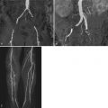
Residual or recurrent coarctation of the aorta is a common complication after either angioplasty or surgical intervention. CMR can not only assess the morphology of the coarctation site but also determine if the lesion is hemodynamically significant, based on the presence of collateral circulation ( Figure 16-3 ).


Feb 1, 2019 | Posted by in MAGNETIC RESONANCE IMAGING | Comments Off on Postoperative Evaluation of Congenital Heart Disease

Full access? Get Clinical Tree

Get Clinical Tree app for offline access