- •
Peripheral arterial disease (PAD) is prevalent, particularly in the elderly.
- •
X-ray angiography has definite risks and limitations, including exposure to radiation and administration of iodinated contrast.
- •
Magnetic resonance angiography (MRA) has emerged as a popular alternative in the diagnostic work-up of patients with PAD.
- •
The most common techniques employed for MRA include time of flight (TOF-MRA), phase contrast (PC-MRA), and contrast-enhanced (CE-MRA) angiography.
- •
TOF-MRA is most commonly used to image the carotid and intracranial circulation.
- •
CE-MRA is less sensitive to artifacts and can be performed rapidly.
- •
The sensitivity and specificity of CE-MRA in PAD is greater than 90%.
- •
Disadvantages of CE-MRA in PAD include timing of gadolinium administration, need for IV access, and concerns regarding gadolinium-induced nephrogenic systemic fibrosis.
Peripheral arterial disease (PAD) is prevalent, particularly in the elderly population. Typical symptoms include claudication and nonhealing ulcers or wounds. Multiple imaging modalities are now available for diagnostic evaluation of the peripheral vasculature. Traditional x-ray angiography has been considered the “gold-standard” technique to define vascular anatomy. However, x-ray angiography has definite risks and limitations, including exposure to radiation and administration of iodinated contrast that may result in allergic reactions and contrast nephropathy. Contrast nephropathy is particularly important in this patient population as many patients with PAD also have renal insufficiency. Therefore noninvasive alternatives, such as magnetic resonance angiography (MRA), have emerged as popular alternatives in the diagnostic work-up of patients with PAD.
Magnetic resonance angiography (MRA) can be accomplished with a variety of techniques. The most common techniques employed include time of flight (TOF-MRA), phase contrast (PC-MRA), and contrast-enhanced (CE-MRA) angiography. Each technique has its distinct advantages, disadvantages, and preferred indications.
TOF-MRA manipulates longitudinal magnetization of stationary spins, thus providing contrast within the vasculature. TOF-MRA is most commonly used to image the carotid and intra-cranial circulation. PC-MRA uses velocity changes, and hence phase shifts to provide contrast within the vasculature. CE-MRA typically utilizes a gadolinium chelate, a T1 shortening agent that, in conjunction with a T1 weighted sequence (typically a conventional or fast gradient echo sequence), leads to high signal intensity of the arteries compared with the surrounding tissue. The two main distinguishing factors in favor of CE-MRA compared with the other two techniques are: CE-MRA is not affected by artifacts caused by in-plane saturation and turbulence, and reduction in examination time (entire study can be completed in less than 15 minutes). Multiple studies have consistently shown the sensitivity and specificity of CE-MRA are greater than 90%. Hence CE-MRA is widely used to evaluate peripheral arterial disease. The disadvantages include problems with timing of gadolinium administration relative to image acquisition, and the need for intravenous access. Recently there also has been concern about gadolinium-induced nephrogenic systemic fibrosis in patients with renal insufficiency.
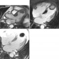
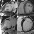
A 49-year-old patient presented with a 5-day history of bilateral calf pain with exertion and at rest. On examination there was severe tenderness on palpation of bilateral lower extremities. Peripheral pulses (femoral, popliteal, anterior tibial, and dorsalis pedis) were normal in amplitude and contour. Laboratory data revealed significantly elevated serum creatine kinase level, white blood cell count, and erythrocyte sedimentation rate. The referring physician was concerned about the integrity of the lower extremity vasculature and ordered a CMR/MRA ( Figure 20-1 ).

Related posts:


Stay updated, free articles. Join our Telegram channel

Full access? Get Clinical Tree


