Key points
- •
Most patients with objective tinnitus, whether pulse-synchronous or not, will have an identifiable and frequently treatable cause.
- •
Venous etiologies of pulse-synchronous tinnitus are the most common.
- •
A thorough diagnostic evaluation must look for causes of abnormal sound production and abnormal sound perception.
- •
Vascular studies must include venous phases, with particular attention to imaging the transverse and sigmoid sinuses.
- •
Subtle sigmoid sinus wall anomalies will only be detected with high-resolution appropriately windowed computed tomography.
Introduction
Surgery for tinnitus can be divided into procedures directed specifically at elimination of tinnitus versus those directed at an independent primary otopathology whose symptoms include tinnitus ( Table 1 ). For the latter, although there may be an independent primary goal for which the surgery is undertaken, tinnitus may be expected to improve secondarily. This article will address both tinnitus-specific and nontinnitus-specific procedures for objective and subjective causes.
| Tinnitus Specific Surgery | Surgery with Secondary Benefit on Tinnitus |
|---|---|
|
|
Tinnitus is defined as the abnormal perception of sound in the absence of an external sound source. Objective tinnitus has traditionally referred to a sound that can be heard by another listener aside from the patient. This term can be misleading, since it is highly dependent on the degree of attention the examiner pays and the tools employed. However, if one defines objective tinnitus as that which, based on its auditory characteristics and other features, is thought to arise from an objective, mechanical sound source, the ambiguity is eliminated. Objective tinnitus can be pulsatile (or, more precisely, pulse-synchronous) or nonpulsatile. Surgical intervention for tinnitus relief is most commonly performed for objective tinnitus.
Using the same rationale for definition, a so-called subjective tinnitus is that which, based on the presumed etiology and pathophysiology, is thought to be caused by a purely electrochemical phenomenon. This type of abnormal sound perception cannot be heard by an objective listener no matter the sensitivity of the listening equipment. Although commonly perceived as a continuous tone, it too can often have rhythmicity, such as that of chirping crickets, although it is only very rarely truly pulse-synchronous or regularly rhythmic. Throughout this article, sounds thought to arise from an objective, mechanical sound source will be referred to as objective tinnitus, and those arising from a purely electrochemical phenomenon will be referred to as subjective tinnitus.
Clinical evaluation
For a detailed discussion of the clinical evaluation of the patient with tinnitus, please refer to Hertzano R, Teplitzky TB, and Eisenman DJ: Clinical Evaluation of Tinnitus , in this issue. To summarize, the focus of the evaluation must be directed both at identifying potentially treatable causes and at assessing the impact of the tinnitus on the patient’s daily activity and well-being. It is important to distinguish early on between objective and subjective causes, because the focus of the evaluation will differ significantly. A thorough history and complete head and neck examination are the critical foundation upon which the further evaluation rests. Complete audiometry will identify and classify any associated, and possibly causative, hearing loss. Most patients with subjective tinnitus have underlying hearing loss. If the audiogram demonstrates symmetric sensorineural hearing loss, the tinnitus itself is perceived bilaterally or with no lateralization, and there are no other associated symptoms or physical examination findings of concern, then no further diagnostic evaluation is required.
The differential diagnosis for pulse-synchronous tinnitus should be divided into causes due to abnormal sound production (eg, transverse sinus stenosis, sigmoid sinus wall anomalies, acquired dural vascular lesions) and those due to abnormal sound perception (eg, third mobile window syndromes) ( Table 2 ). There are also some causes whose pathophysiology is uncertain, such as migraine and Meniere’s disease. If during the initial clinical assessment a particular category is found to be the likely cause for the tinnitus, the remainder of the evaluation will be directed more specifically toward evaluation of this etiology. It is important to note that a significant majority of patients with pulse-synchronous tinnitus will have an identifiable, and often treatable, cause.
| Abnormal Sound Perception | Abnormal Sound Production |
|---|---|
|
|
Patients with a conductive hearing loss on the puretone audiogram must undergo immittance testing. Conductive hearing loss can be associated both with middle ear abnormalities, such as ossicular discontinuity or fixation, and with third mobile window syndromes, such as superior semicircular canal dehiscence. However, whereas patients with the former will typically have absent acoustic reflexes, in third mobile window syndromes, the reflexes should be present. Additionally, patients with a third mobile window will often have symptoms of dizziness with observable nystagmus, evoked by application of positive and negative pressure to the sealed external auditory canal (the so-called fistula test). Vestibular evoked myogenic potentials (VEMP) can also be helpful in distinguishing conductive hearing loss caused by stapes fixation from that of a third mobile window syndrome. With otosclerosis and other causes of stapes fixation, the VEMP responses are typically absent, because the fixed stapes does not provide sufficient mechanical stimulus to vibrate the otolithic organs. With third mobile window syndromes, on the other hand, responses are typically present at abnormally low thresholds.
Radiographic imaging may be indicated following the initial clinical evaluation. Patients with asymmetric sensorineural hearing loss, or chronic, unilateral subjective tinnitus even with symmetric hearing, should undergo diagnostic magnetic resonance before and after administration of gadolinium contrast to look for lesions along the auditory pathway from the labyrinth to the brainstem. Patients with subjective tinnitus associated with other neurologic symptoms, such as vertigo, disequilbrium, headache, or other cranial neuropathies, should be tested with a similar protocol.
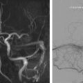
Diagnostic imaging is always indicated for patients with objective tinnitus, although there is controversy about the optimal imaging strategy. For pulse-synchronous objective tinnitus, the authors employ a modified computed tomography (CT) angiography brain protocol, as described in the article on venous abnormalities leading to tinnitus in this issue. Briefly, the contrast injection is delayed slightly to ensure venous enhancement, and the images are acquired with bone windows to allow for identification of fine bony deficiencies. This protocol allows for identification of otic capsule dehiscences causing third mobile window syndromes, sigmoid sinus wall anomalies including subtle dehiscences, soft tissue lesions in the middle ear, and lesions in the jugular foramen. Many indirect signs of acquired dural vascular lesions can be identified as well, such as asymmetrically increased arterial feeders or venous collaterals, transcalvarial channels, inner table scalloping and asymmetric venous attenuation. Asymmetric early venous filling, however, may be missed because of the delayed injection. If this study does not demonstrate a cause for the pulsatile tinnitus, digital subtraction angiography (DSA) is considered. DSA is not recommended as the initial study of choice, because this will not diagnose common causes such as sigmoid sinus wall dehiscence, middle ear abnormalities, or third mobile window syndromes. If DSA is undertaken, it is important to include venous phase images to look for dural venous sinus stenosis and other venous etiologies.
Underlying causes of nonpulse-synchronous objective tinnitus due to presumed middle ear myoclonus are rarely identified with diagnostic imaging. However, most clinicians still feel imaging is indicated to exclude a lesion along the cranial nerve pathways supplying the tensor tympani and stapedius muscles, or those that mediate the afferent pathways of their reflex contraction. This is best done with contrast-enhanced magnetic resonance of the brain with attention to the internal auditory canals and skull base.
Nonspecific treatments (ie, treatments for other indications with secondary tinnitus benefit)
Most patients with severe hearing loss, whether conductive or sensorineural, report an associated, nonrhythmic, subjective tinnitus that likely results from the sensory deafferentation. Consequently, many procedures directed at hearing improvement will secondarily result in an improvement in tinnitus. Most patients who are candidates for cochlear implantation for bilateral, unaidable sensorineural hearing loss also have associated tinnitus. Studies have shown that most of these patients will report an improvement in their tinnitus following implantation. Similarly, preliminary data have suggested that patients with unilateral sensorineural hearing loss and tinnitus who undergo implantation of an osseointegrated bone-conduction device for their hearing loss will also report improvement in their subjective tinnitus. Intratympanic perfusion of steroids has been employed for a variety of idiopathic otopathologies that are commonly associated with tinnitus, including Meniere’s disease, sudden sensorineural hearing loss, and acute labyrinthitis, among other disorders. Studies have shown that many patients undergoing intratympanic perfusion for inner ear diseases associated with tinnitus, particularly patients with Meniere disease, will also report improvement in the associated tinnitus following the procedure.
In addition to a nonrhythmic subjective tinnitus, many patients with conductive hearing loss or conductive hyperacusis will also experience pulse-synchronous tinnitus. Clinical experience suggests that procedures to improve hearing for the former will usually result in improvement in the pulse-synchronous tinnitus, although systematic studies are lacking. Similarly, many, if not most, patients with pulse-synchronous tinnitus associated with superior canal dehiscence syndrome will report improvement in that symptom when canal occlusion or resurfacing procedures are performed. However, there are insufficient data to determine if pulse-synchronous tinnitus alone is an appropriate indication for canal dehiscence surgery.
Finally, surgery for excision of a middle ear or jugular foramen paraganglioma that is also associated with pulse-synchronous tinnitus would be expected to relieve the symptom if the primary lesion is successfully removed.
Related posts:


Stay updated, free articles. Join our Telegram channel

Full access? Get Clinical Tree


