12 Testicular Artery
K.I. Ringe
In older studies and textbooks, the testicular and ovarian arteries were subsumed under the term internal spermatic artery.1–3 Thus, in some studies on variations of these arteries the sex was not mentioned in which the abnormalities were observed.1,4–6 In general, deviations in the origin and course of the testicular and ovarian arteries seem to be comparable. Only the testicular artery is shown in the figures in this chapter, since there are more studies on anomalies of the testicular arteries.7–11 Only a small number of groups have done routine angiographies of the small testicular arteries.12–14 In some rare cases, the testicular arteries arch over the renal arteries first before they turn toward the inguinal canal.1,15 An anastomosis between the artery of the ductus deferens and the testicular artery has sometimes been found along the epididymis.16
12.1 Testicular Arteries Originating Only from the Aorta (83%)
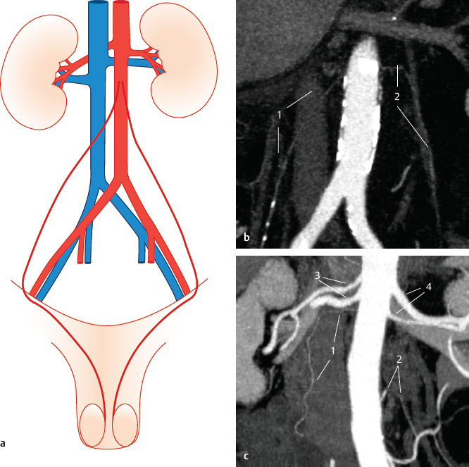
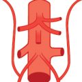
Fig. 12.1 Normal situation as found in textbooks (~68%). On each side, one testicular artery arises from the infrarenal aorta. Schematic (a) and coronal MIP CT images from two patients (b,c). In image c the left testicular artery branches distinctly lower; in addition, two renal arteries are present on both sides. 1 Right testicular artery; 2 left testicular artery; 3 right renal arteries; 4 left renal arteries.
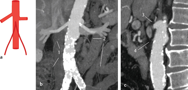
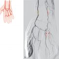
Fig. 12.2 The right and left testicular arteries form a common initial trunk (<0.1%). Schematic (a) and MIP CT (b,c). Coronal (b) and sagittal (c) view of the infra-renal aorta with the right and left testicular artery branching from a common trunk from the aorta. 1 Right testicular artery; 2 left testicular artery; 3 left renal artery; 4 common trunk.

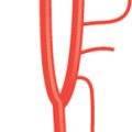
Fig. 12.4 Two testicular arteries on the left side (8%). Schematic.

Fig. 12.5 Two testicular arteries on the right side (4%). Schematic.

Fig. 12.6 Two testicular arteries on each side (2%). Schematic.

Fig. 12.7 Three testicular arteries are found on one side (more often on the left than on the right side) (<1%). Schematic.
12.2 Testicular Arteries Also Originating from the Renal Artery (17%)
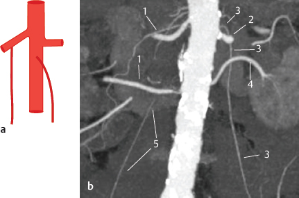
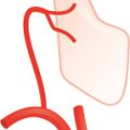
Fig. 12.8 Right testicular artery from the right renal artery (6%). Schematic (a) and coronal MIP CT (b). The CT image shows the right testicular artery arising from the right renal artery and the left testicular artery arising from the suprarenal aorta. Note that two renal arteries are present on both sides. 1 Right renal arteries; 2 left renal artery; 3 left testicular artery; 4 left renal artery; 5 right testicular artery.
Stay updated, free articles. Join our Telegram channel

Full access? Get Clinical Tree


