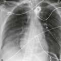
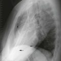
On the normal chest x-ray, we see air in the trachea and proximal bronchi because they are surrounded by the soft tissue (water density) of the mediastinum. In the lungs, however, the bronchi are not visible. The only branching structures visible in the lungs are the pulmonary vessels (water density) surrounded by air.
|
|
|
|
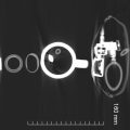
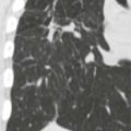
| In Figure 7-1A , the branching pulmonary vessels are visible in the lung. The trachea and proximal main bronchi ( arrows ) are surrounded by mediastinal soft tissue and are visible. The peripheral bronchi are not visible. On computed tomography (CT), the bronchi are normally visible through much of the lung. In Figure 7-1B , a right bronchus ( arrow ) appears tubular (in plane) and a left bronchus ( arrow ) appears circular (perpendicular to plane). Figure 7-1C , a coronal CT reconstruction, shows the distal trachea, carina, and intraparenchymal bronchi in plane ( upper arrow ), perpendicular ( middle arrow ) and oblique ( lower arrow ) as a tube, circle, and oval. | |
|
|
| Figure 7-2A shows a bronchogram with iodinated contrast medium filling normal medial bronchi and dilated lateral bronchi (bronchiectasis). CT has replaced bronchography. In a different patient, Figure 7-2B shows multiple dilated bronchi in cross-section on the left and relatively normal bronchi on the right in cross section (perpendicular). Figure 7-2C is a coronal CT scan that shows the left lower lobe bronchiectasis longitudinally. | |
|
|
|
|
| Straw B is much less visible because there is air inside and outside the thin-walled straw. | |
|
|
|
|
| Figure 7-4 is a scout view of a patient with left lower lobe pneumonia. The bronchi appear as branching black tubes in the consolidated lung behind the heart. In Figure 7-5 , the CT scan shows a right middle lobe air bronchogram. Interstitial thickening elsewhere in the right lung does not give an air bronchogram. | |
| |
|
|
|
|
| Figure 7-6A is a radiograph of a patient with bilateral consolidation. The right upper lobe bronchi are visible (arrows), but the pulmonary vessels are not. Arrows indicate air bronchograms in the right upper lobe. | |
|
|
|
|
| It means the adjacent lung is consolidated. | |
| Figure 7-6B shows extensive air bronchogram in left lower pneumonia. | |
|
|
|
|
|
|
| Patchy peripheral lung consolidation or interstitial disease usually does not cause enough lung opacity to produce an air bronchogram. Conditions that hyperinflate the lungs do not cause air bronchograms. In Figure 7-6A , the right lower lobe and left lung pneumonia are not dense enough to cause air bronchograms. | |
|
|
|
|
|
|
|
|
| In Figure 7-7A , there is no air bronchogram in the collapsed right upper lobe because the bronchi are full of mucous plugs. In this patient, a tumor obstructs the right upper lobe bronchus ( Figure 7-7B , arrow ). | |
|
|
| CLINICAL PEARL: Postoperative left lower lobe atelectasis is frequent and difficult to detect through the heart. Sometimes an air bronchogram seen through the cardiac shadow is the most definitive sign of left lower lobe consolidation. In Figure 7-8 air bronchograms ( arrows ) are visible through the density of the heart. There is also a silhouette sign of the medial diaphragm. | |
|
|
|
|
|
|
|
|
| CLINICAL PEARL: An air bronchogram indicates open airways, strong evidence that the lung disease is not due to an obstructing tumor in a smoker. | |
|
|
|
|
| In Figure 7-6B , the bronchi are normally spaced, whereas in Figure 7-8 they are crowded. | |
|
|
| Bronchiectasis is difficult to diagnose and illustrate on an x-ray. Figure 7-9 shows dilated bronchi ( arrows ) at the lung base. Figure 7-10A shows dilated, thickened bronchi. Bronchi running in the axial plane are tubular ( straight arrows ), and bronchi running across (perpendicular to) the axial plane are circular ( curved arrow ). Figure 7-10B shows dilated bronchi completely filled with secretions in plane ( straight arrows ) and in cross-section ( curved arrow ). | |
| Anagram: Rearrange the letters in DORMITORY to form two words that better define it (answer at end of chapter). | |
Stay updated, free articles. Join our Telegram channel

Full access? Get Clinical Tree


