, Jean-Philippe Bault2, Bernard Benoit3 and Gérard Couly4
(1)
Center of women and fetal imaging, Créteil, France
(2)
Center of fetal imaging Ambroise Paré, Les Mureaux, France
(3)
Princess Grace Hospital, Monaco, France
(4)
Department of maxillo-facial surgery, Necker Hospital, Paris, France
The study of the fetal eye is an important moment in the examination of the face as numerous ocular anomalies are often found in syndromic disorders. Precise knowledge of the of normal semeiology is essential to better understand pathological aspects.
6.1 Embryology Review
Beginning on the 21st day (of pregnancy), neural folds in the diencephaly region invaginate to form gutters that transform into optic vesicles, which then rapidly invaginate again into optic cups. The ectoderm in contact with the vesicle thickens to constitute the lens placode, which in turn invaginations to become the lens vesicle. This then transforms into the lens.
The retina and lens are irrigated by the hyaloid artery (a branch of the ophthalmic artery) which reaches into the ocular globe by the coloboma fissure located on the ventral side of the optic pedicle. If this fissure does not close, there will be coloboma anomalies.
6.1.1 Ultrasound of a Normal Eye
6.1.1.1 First Trimester
The ocular structures are visible as early as the first trimester, in particular the ocular globes, the lens and the orbits. Note that at 13GW, it is possible to visualize a slight posterior thickening of the lens that corresponds to remnants of the primitive vitreous (Fig. 6.1).

Fig. 6.1
The eye during the first trimester
6.1.1.2 Second Trimester
The orbits are perfectly identifiable: in particular in maximum volume mode, which makes it possible to see the various bone components (Fig. 6.2). The ocular globes and the lenses are quite visible; the hyaloid artery appears as a thin line connection the back side of the lens to the retina (it does not persist beyond 28GW) (Fig. 6.3).


Fig. 6.2
Bony orbits (maximum volume mode)
Fig. 6.3
Eye at 22 GW (note the presence of hyaloid arteries)
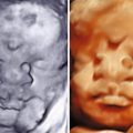
6.1.1.3 Third Trimester
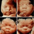
The various eye structures are perfectly visible, as seen in Fig. 6.4, with the pupil being visible on a frontal view (Fig. 6.5).


Fig. 6.4
Eye during the third trimester
Cornea
Pupil
Front chamber
Vitreous body
Lens
Fig. 6.5
Pupil on a frontal view
Pupil
Furthermore, one can see the optic nerve, the ophthalmic artery, the retrobulbar fat (which appears as clearly echogenic) (Fig. 6.6).

Fig. 6.6
Optic nerve, ophthalmic artery, retrobulbar fat
Optic nerve
Ophthalmic artery
Retrobulbar fat
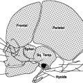
The chiasma opticum can be seen from 20GW using the volume mode. We described the capture in 2006 and the biometrics in 2007 in the journal Ultrasound in Obstetrics and Gynecology (cf. references) (Figs. 6.7, 6.8, and 6.9).


Fig. 6.7
Chiasma opticum at 22, 28, 32 and 34 GW
Fig. 6.8
Chiasma opticum at 22 GW: fetopathological examination (* olfactive bulbs, ° chiasma, Sph sphenoid wings)
Fig. 6.9
Chiasma biometrics
The eyelids and eyelashes are visible from the second trimester (Fig. 6.10).

Fig. 6.10
Eyelids and eyelashes (yellow arrow)
It is possible to evaluator the fundus (virtual) using volumetric capture using the technique that we described in 2008 in the journal Ultrasounds in Obstetrics and Gynecology (cf. References) (Figs. 6.11 and 6.12).


Fig. 6.11
Capturing the eye fundus
Fig. 6.12
Fundus at 16 GW (note the slight depression corresponding to the insertion of the hyaloid artery) at 25 GW and 36 GW
It is possible to measure the inter-orbital distance. In France, we tend to use the measurement from the middle of one orbit to the middle of the other (Fig. 6.13), the relationship between the IOD/BPD is constant: 0.47 at 22 GW and 0.42 at 32 GW.

Fig. 6.13
Interorbital distance measurement
IOD
A simple technique is that of the “virtual orbit”: if the two eyes are separated a normal distance, it is possible to put just one “virtual orbit” in between the two normal orbits (Fig. 6.14).

Fig. 6.14
Virtual orbit
When making a diagnostic study, one will use various published tables, in particular those regarding measurements regarding the orbits, the ocular globes and the lenses.
In addition, an attentive study will make it possible to visualize ocular movements. It should be noted that strabismus, particularly when divergent, is often observed in the fetus.
6.1.2 Main Aspects of Ocular Pathologies
6.1.2.1 Dacryocystocele
This is a cyst in the lachrymal canal that can be seen on an ultrasound as a small swelling in the internal corner of the eye. On an axial view, this cyst appears as an anechogenic formation inside and to the front of the eye. It is sometimes possible to reveal a small lithiathis in the cyst (Fig. 6.15). In the majority of cases, these dacryocystoceles are easily resolvent after birth or even in utero (Fig. 6.16).