The Many Combinations of MRI
Introduction
Thus far we have studied a vast array of MRI sequences that seem somewhat related to one another, but also, quite different. The pulse sequences that we have looked at are the basic sequences used in MRI; however, there are many more combinations of sequences that can be performed and in this chapter we will look at how these interrelate.
Basic Building Blocks
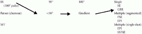
The basic elements needed to build an MRI pulse sequence can be simplified into four components: (i) prep pulse (optional), (ii) radio frequency (RF) pulse, (iii) refocusing mechanism, and (iv) readout (Table 31-1). This is obviously a fairly simplified approach and omits certain items such as the phase-encode step, which is in every sequence. It also puts in an artificial distinction between the gradient refocusing mechanism in gradient-echo (GRE) sequences and the frequency readout, which are being performed simultaneously. Additionally, the refocusing 180° RF pulses in the fast spin-echo (FSE) technique are important both for refocusing and for making multiple readouts. Regardless of the few shortcomings, this approach is useful for understanding the new sequences that the manufacturers are offering on their magnets.
Prep Pulse (Optional). The prep pulse comes temporally before the other three elements, but it is also the one component that is optional. When a prep pulse is used in a sequence, it can actually have an important part to play in the appearance of the image as the root pulse sequence. There are three basic types of prep pulses: (i) 180° RF inversion pulse, (ii) a fat saturation (or chemical saturation) pulse (usually 90° RF pulse), and (iii) a magnetization transfer (MT) pulse. All three of these pulses have different characteristics and properties, with resultant different effects. We have discussed all these pulses in prior chapters; however, it is important to note that any of these can be used with any pulse sequence to achieve a desired effect on the image.
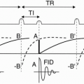
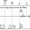
For instance, a 180° RF pulse is used in both STIR and FLAIR imaging with the only variables being the TI, TR, and TE (conventional inversion recovery [IR] and fast IR pathways are shown in Table 31-2; a novel application is seen in Fig. 31-1). The 180° RF pulses can also be used to suppress flow such as in a double inversion recovery (DIR) (or black-blood) technique that is useful in cardiovascular imaging. This technique uses a nonslice selective 180° pulse followed by another 180° pulse that is slice selective. This idea can be carried out further by adding a third 180° pulse that nulls fat (known as a triple IR or fat-saturated blackblood technique—really just a combination of STIR and DIR) (Fig. 31-2). An inversion pulse is also useful in cardiac imaging for identification of infarcted myocardium in delayed enhancement imaging. In this technique, gadolinium is given and the heart is imaged 10 to 20 min later using an IR–GRE technique with an inversion
time of 200 to 300 msec to null normal myocardium (Table 31-3 and Fig. 31-3).
time of 200 to 300 msec to null normal myocardium (Table 31-3 and Fig. 31-3).
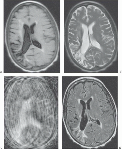 Figure 31-1. A: An SSFSE FLAIR image of the brain in an uncooperative patient. There are corresponding SSFSE (B), T2 (C) with marked motion, and later FLAIR (D) images when the patient was more cooperative. There are periventricular white matter ischemic changes and an old right parietal stroke. This example demonstrates a novel approach for newer fast imaging sequences that can be used in uncooperative patients. |
Table 31-1 The Many Combinations of MR | |||||||||||||||||||||||||||||||||||||||||||||||||||||||
|---|---|---|---|---|---|---|---|---|---|---|---|---|---|---|---|---|---|---|---|---|---|---|---|---|---|---|---|---|---|---|---|---|---|---|---|---|---|---|---|---|---|---|---|---|---|---|---|---|---|---|---|---|---|---|---|
| |||||||||||||||||||||||||||||||||||||||||||||||||||||||
Table 31-2 Inversion Recoverya | ||||||||||||
|---|---|---|---|---|---|---|---|---|---|---|---|---|
| ||||||||||||
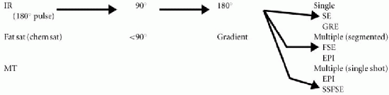
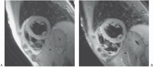 Figure 31-2. A: Short-axis DIR (double inversion recovery) or black-blood technique. B: The same slice using the TIR (triple inversion recovery) technique—notice the nulling of the fat on the TIR versus the DIR. |
| ||||||||||||
Related posts:
Stay updated, free articles. Join our Telegram channel

Full access? Get Clinical Tree


