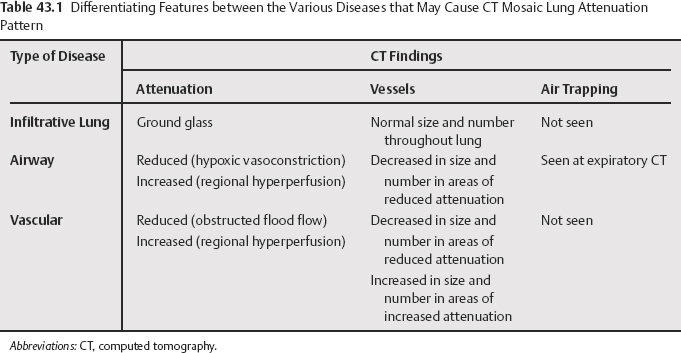
43 A mosaic pattern of attenuation, with patchy areas of increased and decreased attenuation, is nonspecific and may be seen on thin-section computed tomography (CT) of lungs when various infiltrative lung, airway, or vascular diseases are present (Table 43.1). When this pattern is caused by regional differences in perfusion due to vascular diseases, it is also known as mosaic perfusion or mosaic oligemia.
The Mosaic Pattern of Lung Attenuation

Differential Diagnosis of Mosaic
Stay updated, free articles. Join our Telegram channel

Full access? Get Clinical Tree


