- 4
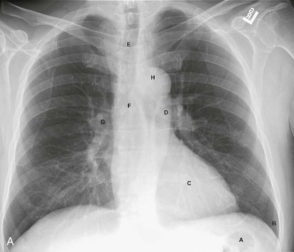
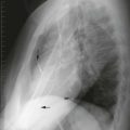
Test yourself by labeling the radiographs in Figures 3-2A and 3-2B .
|
- 4
- A.
gas in splenic flexure
- B.
costophrenic sulcus (angle)
- C.
heart
- D.
descending aorta
- E.
trachea
- F.
carina
- G.
hilum
- H.
aortic knob
- J.
ascending aorta
- K.
right diaphragm
|
- A.
_____________
- B.
_____________
- C.
_____________
- D.
_____________
- E.
_____________
- F.
_____________
- G.
_____________
- H.
_____________
- J.
_____________
- K.
_____________
|
heart
descending aorta
costophrenic sulcus (angle)
gas in splenic flexure
right diaphragm
aortic knob
trachea
carina
hilum
ascending aorta
|
To maximize your accuracy, you must have an organized search pattern. Start reading every radiograph—chest or otherwise—by scanning the areas of least interest first, working toward the more important areas. You are less likely to miss secondary but important findings this way. For the chest x-ray, start in the upper abdomen, then look at the thoracic cage (soft tissues and bones), then the mediastinal structures, and finally the lung. Look at each lung individually, then compare left lung and right lung. A helpful mnemonic is Are There Many Lung Lesions (ATMLL)? |
- 5
Arrange the following in viewing sequence:
|
- 5
- A.
abdomen [A]
- B.
thorax and soft tissue [T]
- C.
mediastinum [M]
- D.
lung—unilateral [L]
- E.
lung—bilateral [L]
|
- A.
_____________
- B.
_____________
- C.
_____________
- D.
_____________
- E.
_____________
|
mediastinum
lung—unilateral
abdomen
lung—bilateral
thorax and soft tissue
|
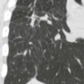
Abdomen: In Figure 3-3A , start in the right upper quadrant (*) and scan across the upper abdomen several times. Normal gas-containing structures are the stomach and the splenic flexure of the colon. The liver is always visible. The hepatic flexure and the spleen may be visible. |
- 6
Scan the abdomen in Figure 3-3B .
|
- 6
- A.
stomach bubble
- B.
splenic flexure (colon)
- C.
liver
- D.
right diaphragm
|
- A.
The gas collection just below the heart = _____________.
- B.
The gas collection lateral to A = _____________.
- C.
The homogeneous density below the right diaphragm = _____________.
- D.
The higher diaphragm is the _____________.
|
liver
stomach bubble
splenic flexure (colon)
right diaphragm
left diaphragm
hepatic flexure
|
CLINICAL PEARL: Upper abdominal disease (subphrenic abscess, perforated viscus, pancreatitis, and cholecystitis) may mimic lung disease clinically. Similarly, basilar lung disease (pneumonia, pleurisy) may mimic upper abdominal disease. This is real! You’ll see! |
Mediastinum: An organized search of the mediastinum is complicated because there are multiple overlapping structures. Start with a global look at the mediastinum for contour abnormalities (i.e., focal or diffuse widening). Figures 3-4A and 3-4B show three rapid searches of the mediastinum: A = for the trachea and carina; B = for the aorta and heart; C = for the hilum. |
- 7
Test yourself on Figure 3-4C by identifying the following structures:
|
- 7
- 1.
trachea
- 2.
carina
- 3.
aortic knob (arch)
- 4.
ascending aorta
- 5.
descending aorta
- 6.
heart
- 7.
right hilum
|
- 1.
_____________
- 2.
_____________
- 3.
_____________
- 4.
_____________
- 5.
_____________
- 6.
_____________
- 7.
_____________
|
aortic knob (arch)
carina
heart
trachea
right hilum
ascending aorta
descending aorta
|
Note that the left hilum is normally slightly higher than the right. Just to confuse, the right diaphragm is normally slightly higher than the left. |