MRI: Best to evaluate extent. Mass isointense/slightly hyperintense to normal white matter on T1 and T2 and shows prominent CE. Causes fusiform enlargement of nerve, which is buckled and kinked.
One-third in the setting of NF1 and characteristically bilateral.
Hemangioma
CT/MRI: usually extraconal but some lesions intraconal.
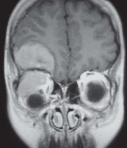
CT/MRI: unilateral or bilateral uveoscleral thickening with enhancement, an enhancing retro-ocular mass, or enlargement of extraocular muscles, tendons, and lacrimal gland.
Idiopathic disorder with inflammatory, lymphoid infiltration of intraorbital tissues.
Lymphoma and metastases
CT/MRI: Lymphoma: diffuse infiltration of entire intraconal region. Shows slight to moderate enhancement.
Lymphoma best imaged with MRI.
Metastases: neuroblastoma/rhabdomyosarcoma, usually also with extraconal and orbital wall involvement.
Hematoma
CT: Hyperdense if acute. CT best for evaluating fractures.
CT/MRI: May be primary or secondary. Usually intraconal or involving muscle but may extend extraconal and intracranial. MRI best for extent: T1 iso-/hypoin-tense relative to muscle and T2 hyperintense. Degree of enhancement varies. Enhanced scans in coronal plane best for evaluating intracranial extension.
Usually embryonal subtype.
Fig. 4.126a, b T1-weighted sagittal oblique pre- (a) and postcontrast (b) MRI: a right optic nerve glioma with fusiform enlargement of the nerve, which shows marked enhancement postcontrast.Fig. 4.127 Axial postcontrast CT shows enhancing left intraconal mass with anteromedial, extraconal extension. The biopsy proved rhabdomyosarcoma.Fig. 4.128 T2-weighted coronal MRI shows large hyperintense intraconal rhabdomyosarcoma causing marked proptosis.
CT/MRI: well-demarcated, anterior extraconal mass with fatty, fluid, or mixed contents. Majority superolateral related to frontozygomatic suture. CT shows osseous remodelling ± fine calcification in cyst wall.
MRI if features are not diagnostic or suspect deep extension into sinuses, masticator space, or intracranially.
Usually complex with cystic and solid elements, composed fat/fluid/calcium and soft tissue. Often marked expansion of the orbit and displacement of the globe.
Benign but cause severe progressive proptosis in infancy. Transspatial.
Capillary hemangioma
MRI is imaging modality of choice to show extent especially if suspected intracranial extension. Use enhanced T1 and T2, axial, and coronal planes. T1 iso- or hypointense, showing diffuse intense enhancement postcontrast. T2 heterogeneous/hyperintense with frequent flow voids. MRA unhelpful as vascular component is at capillary level.
CT: slightly hyperdense homogeneous mass with intense enhancement.
Usually present before 6 mo. Only imaged if symptomatic or fail to involute.
Lymphatic, venous, and venolymphatic malformations
MRI best to show extent; use fat-saturated axial and coronal sequences. See multilocular, transspatial, variably enhancing mass. Fluid-fluid levels with signal corresponding to age of blood products. Pure lymphatic malformation is nonenhancing. Venolymphatic variable.
Present in childhood with progressive, slow-growing mass. Usually extraconal but often transspatial.
Include brain imaging for associated intracranial abnormalities especially in venolymphatic malformations.
CECT: thickening and edema of orbital soft tissues representing cellulitis and/or phlegmon. Low-density, rim-enhancing area represents subperiosteal abscess. Usually extraconal but can extend intraconally. Procure axial and coronal images.
Need brain imaging (CT or MRI) to evaluate intracranial complications of sinusitis. MRI more sensitive.
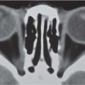
Fig. 4.129 T2-weighted axial MRI shows hyperintense medial extra-conal mass related to frontoethmoidal suture, which is characteristic of a dermoid cyst.Fig. 4.130 T1-weighted coronal MRI shows a large right extraconal mass with extension into right maxillary sinus and face. It is hyperin-tense on T1 with focal hypointensity in keeping with fat and calcification, which is characteristic of a teratoma.Fig. 4.131 T2 coronal MRI shows an intermediate signal intensity, inferior, extraconal mass, with a second mass seen centered on right maxillary sinus; biopsy proved it to be lymphoma.Fig. 4.132 Orbital cellulitis/subperiosteal abscess. Peripheral rim-enhancing medial subperiosteal collection displacing the left globe laterally on coronal postcontrast CT. Note also the opacification of the left ethmoid and maxillary sinuses.
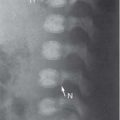
Radiograph/CT: show defect or destruction often not confined to orbits. CT/MRI shows adjacent soft-tissue mass and exophthalmos.
Most common causes are neuroblastoma, hematological malignancy, rhabdomyosarcoma, and Ewing sarcoma.
Extension from lacrimal gland/tumor
Radiograph/CT/MRI: Localized wall expansion adjacent to tumor. Lesions have a sclerotic rim.
Spread of adjacent infection
CT: usually subperiosteal abscess but if longstanding may cause destruction of orbital wall.
Wall involvement usually due to aggressive pathogen particularly aggressive fungal sinus infections such as mucormycosis and aspergillosis (see Table 4.35).
Adjacent tumor
CT: ethmoid or maxillary antral tumors invade by direct extension.
Mucocoele
Radiograph/CT: Bony expansion of adjacent sinus, which protrudes into orbit. Sinus may be sclerotic if chronically infected. Contains nonenhancing soft tissue.
Present with unilateral proptosis. Most common is frontal sinus with palpable mass in superome-dial orbit.
LCH
Radiograph/CT: Single or multiple lytic lesions with beveled edges and adjacent soft-tissue mass. Often have exophthalmos.
MRI needed if sphenoid bone involved and present with diabetes insipidus.
Need skeletal survey to look for other bone lesions.
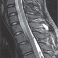
Fig. 4.133 T1-weighted coronal postcontrast MRI shows enhancing right extraconal mass with intracranial extension characteristic of n euroblastoma metastases.
Only gold members can continue reading. Log In or Register to continue