The Pediatric Vertebral Column
The vertebral column consists of 7 cervical, 12 thoracic, and 5 lumbar levels, as well as 5 sacral segments and a few coccygeal levels. The dens, eventually attaching to the body of C2, replaces what would have been the body of C1.
Variants. In a series of 100 radiographs of patients aged between 4 months and 19 years, five complete lumbar vertebrae (without ribs) were present in 84% of cases, four in 5%, and six in 6%. A transitional vertebra with one side sacral and one side lumbar, or else a rib on what would be one side of L1, made up the remaining 5%. These variations are effects of hox genes and may also be seen in the VATER (vertebral, anal, cardiac, tracheoesophageal, radial or renal) association.
Normal findings. A vertical midline cleft is normal in infants; the narrow interface lucency projects over the center of the vertebral body on frontal projections. The cleft progressively ossifies (fuses) caudocranially in thoracic levels in the first year of life. This line can persist as a laminar cleft at C1 and C7, and at the lower thoracic column; it then may serve as a marker to identify individuals. At L5, the cleft often does not fuse until age 10; thereafter, it is known as spina bifida occulta.
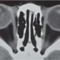
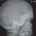
In neonates ( Fig. 4.192 ) on the lateral image, the prenatal bone is relatively dense compared to the bone laid down in the first weeks of life, just adjacent to the denser end plates, which are cartilaginous zones of provisional calcification. The anterior arch of C1 may not ossify during the first year of life, but may be present at birth. The horizontal Hahn fissure, or vascular canal, causes the anterior midbody notch of the biconvex vertebral body, and gradually fades after the neonatal period. Similarly, the neurocentral synchondrosis (see Figs. 4.192 and Fig. 4.268 ) may normally be unossified in early infancy. Anterior steplike projections extend out from the vertebral bodies of school-aged children. Ring apophyses appear in the unossified corner areas at approximately 10 years of age. Thoracic vertebral bodies may have mildly decreased height anteriorly and appear wedged. In the first year of life, the dens is connected to C2 body by a nonossified synchondrosis.
On frontal views of the chest, the cervical spine area often seems quite abnormal in infants, merely due to projection.

Related posts:


Stay updated, free articles. Join our Telegram channel

Full access? Get Clinical Tree


