The Pediatric Vertebral Column: Anomalies of the Spinal Canal and Neural Arches
10.1055/b-0034-87914
The Pediatric Vertebral Column: Anomalies of the Spinal Canal and Neural Arches
Narrowing of the Spinal Canal (Spinal Stenosis)
Narrowing of the spinal canal may be generalized and thus is an indication of slowed enchondral bone formation in bone dysplasias (Fig. 4.239).
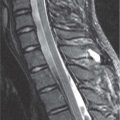
Fig. 4.239 MRI of lumbar spinal stenosis. Sagittal T1-weighted MRI in a 16-year-old with Ehlers-Danlos syndrome. The canal is narrowed in association with shorter than normal lumbar pedicles; the disks bulge somewhat. Note the reduced amount of canal fat, especially compared to Fig. 4.234.
Narrowing of the lumbar canal both coronally (progressive narrowing of the space between pedicles at each lumbar level as one goes inferiorly). Posterior margin of lumbar bodies are notably concave. Cervical and thoracic canal also narrow; occasionally severe in upper cervical region.
Minimal lumbar disk herniation can cause severe symptoms. Occasionally, the cervical canal narrowing may cause respiratory difficulty. Narrowed jugular foramina may cause dilated ventricles; foramen magnum also relatively small.
Hypochondroplasia
Less pronounced changes than achondroplasia. The decrease downward in distance between pedicles is less evident or distance may be constant downward.
Allelic to achondroplasia. Head circumference at least fiftieth percentile.
Idiopathic lumbar spinal stenosis
Isolated variation. Perhaps just the lower range of the bell-shaped curve of spinal canal area.
May be associated with back pain from minor disk protrusions.
Metatropic dysplasia
Cervical spinal stenosis frequent.
Develop kyphoscoliosis during childhood. May have dens hypoplasia.
Consequence of fractures of bodies or arches
Oblique plain images may help define, but CT and MRI are definitive.
Associated cord damage.
Intraspinal exostosis
A rare and unfortunate localization in multiple cartilaginous exostoses.
CT and MRI to define.
Hurler syndrome and other heteroglycanoses
Thickened meninges narrow the intraspinal space.
Hypertrophied ligamentum flavum
CT or MRI to define.
Sirenomelia
Single midline lower extremity with one or two sets of limb bones in it.
Other skeletal conditions with narrowing of the spinal canal include the following:
Atlantoaxial subluxation
Down syndrome
Cartilage hair hypoplasia
Kniest disease
Alagille syndrome
– Multiple stippled epiphyses
– Dyssegmental dysplasia
Diastrophic dysplasia
Acrodysostosis
Gordon syndrome (with narrowed disk spaces)
Acromesomelic dysplasia
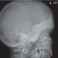
Fig. 4.240a, b Achondroplasia in a 14-year-old girl. (a) Caudally decreasing lumbar transverse interpedicle distance. (b) Classic myelogram: marked spinal stenosis with posteriorly concave bodies.
Only gold members can continue reading. Log In or Register to continue