The Pediatric Vertebral Column: The Spinal Canal and Its Contents


10.1055/b-0034-87917

The Pediatric Vertebral Column: The Spinal Canal and Its Contents


Localized Spinal Stenosis Caused by Bony Changes


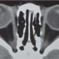
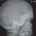
See page 442 for generalized stenosis. Standard investigation includes radiographs in at least two projections and additional work-up with CT. MRI is the method of choice for evaluating the contents of the canal (as well as the medullary portions of bony elements).


Table 4.107 Localized spinal stenosis caused by bony changes

Diagnosis


Findings


Comments


Spondylolisthesis


Posterior displacement of the lower involved level.


Mostly L5-S1; less often L4-L5.


Narrowing more pronounced in grades III and IV. Associated spondylolysis.


Displaced fracture fragments


CT for bony detail; MRI for cord involvement and damage.


Posterior hemivertebra


Often protrudes back into the spinal canal.


Retrolisthesis and kyphosis at that level.


Malformations


For example, Klippel-Feil syndrome.


Disk prolapse


Fig. 4.278a, b


MRI to define.


Causes include spondylodiskitis, calcified nucleus pulposus, and trauma.


Severe scoliosis


Curvature in any direction can narrow the spinal canal and compress nerve roots.


MRI prior to surgery.


Tumors of the vertebral body or arch


Gibbus


Sharp, severe kyphosis.


Classically, in tuberculosis—may increase rapidly.


Achondroplasia


Short pedicles and laminae in cervical column.


May happen in other dysplasias with enchondral slowing, or even from isolated shortened arch elements.

Fig. 4.278a, b Spondylodiskitis in a 5-year-old child. MRI in acute spondylodiskitis of L4-L5. (a) T1-weighted image: decreased signal intensity of both vertebral bodies and poor definition of the disk space. (b) T2-weighted image: increased signal intensity of the narrowed disk space with posterior prolapse and compression of the dural sac (arrow), as well as protrusion into the body of T5.


Masses Within the Spinal Canal


The cervical and lumbosacral canal are normally relatively wide; according to Petersson and Harwood-Nash, the dural sac is significantly wider in approximately 4% of myelograms in children. CT and especially MRI are the diagnostic modalities of choice for those cases that require further diagnostic work-up, whether an intraspinal mass is suspected or if neurologic symptoms occur without bony changes on conventional radiography.


Table 4.108 Masses within the spinal canal

Diagnosis


Findings


Comments


Tumors: extradural and intradural, extramedullary, and intramedullary


Fig. 4.279, p. 478


MRI: low signal on T1 and high on T2 (except fat- or cartilage-containing tumors).


Drop metastases result from astrocytoma and cerebellar tumors such as medulloblastoma.


Lipoma


Fig. 4.280a, b, p. 478


MRI: high signal intensity on T1.


Hematoma


MRI: can appear as high signal in the early phase.


Other sequelae of trauma: necrosis, atrophy, syringomyelia, edema.


Edema of cord


MRI: high signal on T2, low on T1.


Narrowing of surrounding spaces.


CSF after failed lumbar puncture


Evident on US and MRI (and CT) around cord.


Typically regresses in a few days. Usually not blood.


Arachnoiditis


MRI: irregular structures of soft tissue within the dural sac (soft-tissue density on CT).


This was relatively common after administration of oily myelographic contrast medium. This is no longer the case.


Meningeal thickening in MPS


Soft-tissue thickening between the dural sac and the bony vertebral structures.


Compression of the cervical cord has been described in MPS I, I-H/S, I-S, II, IV, and VI.


NF1


Posterior scalloping of vertebral bodies.


Vascular malformations


MRI: signal dropout on T1 and T2, except in small vessels with low-flow velocity.


MRA: artery of Adamkiewicz may be a feeder.


Increased fat deposition in steroid therapy


MRI characteristics the same as lipoma.


Can cause neurologic symptoms.

Fig. 4.279 Astrocytoma expanding spinal canal in 6-year-old boy. The mass at the C1–C6 is seen to expand the spinal canal on T1 sagittal MRI.
Fig. 4.280a, b Intraspinal lipoma, part of lipomeningocele. This 4-year-old child has had release of tethered cord and portions of the lipomeningocele. Note the extension of the fatty elements forward to indent and narrow the lowest lumbar bodies.


Anomalies of the Spinal Cord With or Without Bony Abnormality


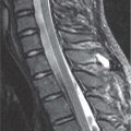
Decreased motion, usually associated with cord tethering, can be determined with US in infants. It is best seen in the first few months of life. Thereafter, MRI shows cord location in sagittal and cranial caudal directions. The conus medullaris does not normally terminate below L3 at any age. Spinal cord tumors are best shown on MRI.


Table 4.109 Anomalies of the spinal cord with or without bony abnormality

Diagnosis


Findings


Comments


Tethered cord


Common in spinal dysraphism. The conus attachment is abnormally low and the cord lacks the normal pulsating motion. Association with meningomyelocele and/or lipoma.


Clinical signs: skin changes, neurologic symptoms, orthopedic abnormalities in the lower limbs such as pes cavus, bladder dysfunction, kyphoscoliosis.


MMC, meningocele, myelocele, lipo-MMC


Spinal dysraphism. MRI: frequently tethered cord with lipoma and high signal of T1.


Severe neurologic deficits. Associated with Chiari type II malformation.


Diastematomyelia


Fig. 4.281a, b


Fig. 4.241, p. 444


Often a bony spur on radiography or CT. Split cord, conus, and/or filum terminale.


May be associated with hydromyelia, scoliosis, Sprengel deformity. Neurologic symptoms are often minimal.


Syringomyelia, hydromyelia


On radiograph may be widening of the spinal canal. The syrinx is positioned centrally and has low T1 signal.


Occurs after trauma and surgery as well as dysraphism, particularly following shunting of a MMC and with Chiari type II malformation.


Spinal cord injury without radiologic abnormality (known as SCIWORA)


After trauma, damage to the cord (shown on MRI) without abnormality of bone on plain radiographs.


Neurologic examination is important to suspect it, but also consider depending on mechanism of injury.

Fig. 4.281a, b Diastematomyelia. (a) Vertical bone spur at L3 level (arrow) and right-sided fused neural arch laminae of L2 and L3. (b) Axial MRI: split cord (C) at the level of C3. The arrow indicates the low signal bone spur.


Changes in the Disk Spaces


Table 4.110 Narrowed disk spaces

Diagnosis


Findings


Comments


Forme fruste of vertebral body fusion


A step along the way to vertebral body fusion is a narrower than normal disk space.


A possible part of VATER association. A hox gene effect.


Spondylitis-diskitis


Disk narrowing may be the first radiographic finding (but not seen in osteomyelitis limited to the body).


Early scintigraphic diagnosis: increased activity in the adjacent vertebral bodies. Well demonstrated with MRI. Disk material may occasionally protrude through the end plates or posteriorly into the spinal canal.


Scheuermann “disease”


Thoracic kyphosis, narrowed disk spaces, irregular end plates, Schmorl nodes.


May have similar configuration in anterior disk prolapse.


Acute Schmorl nodes


Ill-defined margin of the end plate defect. Later sclerosis of the rim.


MRI (or CT) if needed to confirm.


Trauma


Acute traumatic prolapse of disk into spongiosa uncommon in children.


Demonstrated by CT. Sclerosis of edges later.


JIA


Disk space narrowing precedes full fusion.


Associated arch fusions and areas of erosion in cervical spine.


Spondyloepiphyseal dysplasias, Morquio disease, Kniest disease, Cockayne syndrome


Narrowed disk spaces with irregular end plates may be seen in many syndromes.


Table 4.111 Widened disk spaces

Diagnosis


Findings


Comments


Normal variant


The normally flat disk may be centrally biconvex.


Also may occur after hyperextension injury.


Platyspondyly


Lower than normal height of vertebral bodies often is associated with taller than normal disks.


See Tables 4.64 and 4.65 .


Osteoporosis


Weaker than normal end plates allow disks to approach a more spherical shape.


Codfish vertebra configuration.


Other acquired causes of decreased vertebral body height


Sometimes, the disks become taller and sometimes they maintain normal height.


Table 4.112 Disk calcification

Diagnosis


Findings


Comments


Calcification in the nucleus pulposus, solitary or multiple


Fig. 4.282


Most common in the cervical spine, where most commonly associated with pain. Adjacent end plates may be irregular. May prolapse anteriorly or posteriorly.


Persists or resolves spontaneously. Increased incidence in hyperparathyroidism, hypervitaminosis D, homocystinuria, and spondyloepiphyseal dysplasia tarda.


Ochronosis


Disk calcification or ossification; narrows disks.


Onset usually in adulthood. Especially frequent in Czech Republic.

Fig. 4.282 Cervical disk calcification (arrows) in an 8-year-old boy. Intermittent pain and anterior prolapse.


Table 4.113 The vacuum phenomenon

Diagnosis


Findings


Comments


Vacuum phenomenon


In general, air or vacuum with a disk is associated with degenerative disease.


Rare in children. Occurs after trauma. Flexion extension series may reveal a vacuum.

Only gold members can continue reading. Log In or Register to continue

Stay updated, free articles. Join our Telegram channel

Jul 12, 2020 | Posted by in PEDIATRIC IMAGING | Comments Off on The Pediatric Vertebral Column: The Spinal Canal and Its Contents

Full access? Get Clinical Tree

Get Clinical Tree app for offline access