27 The Profunda Femoris Artery
T. Rodt, M. Lee
Various branching patterns of the common femoral artery into the superficial femoral artery and the profunda femoris artery can be found.1–14
27.1 Origin and Branching Pattern

Fig. 27.1 Common trunk supplies the profunda femoris, the medial, and lateral circumflex arteries of the femur (58%). Schematic (a), posteroanterior (PA) DSA (b), and 3D-VR CT images in the right anterior oblique (RAO) view (c), anteroposterior (AP) view (d), and left anterior oblique (LAO) view (e).

Fig. 27.2 The medial circumflex artery is a direct branch of the femoral artery (18%). Schematic (a), axial MIP CT (b), and 3D-VR CT images in the AP view (c), LAO view (d), and left lateral view (e). 1 Lateral circumflex artery; 2 profunda femoris artery; 3 common femoral artery; 4 medial circumflex artery.

Fig. 27.3 The lateral circumflex artery is a direct branch of the femoral artery (15%). Schematic (a), axial MIP CT (b), and 3D-VR CT images in the RAO view (c), AP view (d), and LAO view (e). 1 Common femoral artery; 2 profunda femoris artery; 3 medial circumflex artery; 4 lateral circumflex artery.

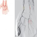
Fig. 27.4 Both circumflex arteries originate directly from the femoral artery (4%). Schematic (a), coronal MIP MR (b), and PA DSA (c). 1 Common femoral artery; 2 medial circumflex artery;
3 lateral circumflex artery;
4 profunda femoris artery.

Fig. 27.5 Same as Fig. 27.1 or Fig. 27.2, but a major branch of the lateral circumflex artery comes from the femoral artery (3%). Schematic (a), axial MIP CT (b,c), and RAO projection 3D-VR CT (d).
1 Lateral circumflex artery;
2 common femoral artery;
3 medial circumflex artery;
4 profunda femoris artery;
5 accessory lateral circumflex branch.
Related posts:


Stay updated, free articles. Join our Telegram channel

Full access? Get Clinical Tree


