The Temporal Bone: Congenital Anomalies
During embryonic development, the external and middle ear are derived from the first and second branchial groove and pouch. The inner ear is derived from surface ectoderm. Its development starts in the third week and is completed around the twelfth week. In the majority of children with congenital sensorineural hearing loss, no anatomic anomaly is demonstrated. Combined anomalies of external, middle, and inner ear occur in approximately 10% of patients.
In external ear atresia, it is important to note whether the atretic plate is composed of soft tissue or bone. The extent of ossicular chain malformation can differ from a fusion of the mallear head and the incudal body to a small clump of malformed chain. Bilateral external ear canal atresia is often associated with a syndrome, while unilateral atresia is usually nonsyndromal.
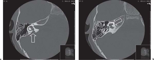
The lateral semicircular canal develops last. In malformations of the semicircular canals, the lateral canal is most commonly affected.
The vestibular aqueduct is widened when its diameter is larger than the diameter of the posterior semicircular canal, which corresponds with a diameter at its midpoint of 1.5 mm. It is almost always associated with cochlear modiolar deficiencies.
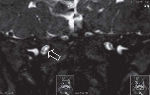
Absence of the cochlear nerve is rare. The internal auditory canal can be narrowed, but it can also occur with an internal auditory canal of normal diameter. On heavily T2-weighted images perpendicular to the internal auditory canal, the anteroinferiorly located cochlear nerve is absent.
Stay updated, free articles. Join our Telegram channel

Full access? Get Clinical Tree


