The Thoracic Aorta


Key Points


  • The chest radiograph is useful to prompt consideration of aortic pathologies but is not able to reliably offer a final diagnosis for most, which is reserved for more advanced imaging modalities such as computed tomography (CT), magnetic resonance imaging (MRI), transesophageal echocardiography (TEE), and aortography.


  • The silhouettes of the aorta should be understood in detail, as should the imaging features on radiography, and by other modalities, of specific acquired, congenital, and repaired aortic pathologies.


Issues Concerning Chest Radiographic Imaging of the Thoracic Aorta


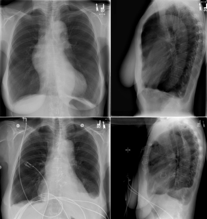
It is important to remember that (1) the chest radiograph does not image the aorta comprehensively and (2) abnormalities of the aorta evident on the chest radiograph represent substantial pathology that is often clinically important and sometimes life-threatening. Therefore, to not overlook major aortic pathology when reading a chest radiograph, it is imperative to scrutinize aortic contours and appearance ( Fig. 7-1 ). The aorta is evident on the chest radiograph wherever the air-filled lung is in contact with it and there is a silhouette to evaluate.


Figure 7-1


There is a markedly abnormal contour to the left of the heart, which is actually of the descending aorta from which a very large sarcoma of the aorta is growing into the left chest cavity.


It is critical to recall that major aortic pathologies, such as dissection, may occur with a normal or near-normal chest radiograph. A final diagnosis for aortic pathology requires a more advanced and comprehensive test such as CT, MRI, TEE, or aortography.


Review of Silhouettes of the Aorta


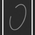
The aortic root begins behind the pulmonary artery at the level of the aortic valve and lies within the pericardium. The root is surrounded by soft tissue (the pulmonary artery in front, the left atrium inferiorly, the pericardium, and pericardial and mediastinal fat) and the aortic valve is usually not evident unless it is either heavily calcified or prosthetic; therefore, the aortic root cannot be localized accurately on the chest radiograph on either the frontal or lateral radiographs. Marked dilation ( Graphic 7-1 ) of the root, which usually occurs concurrently with dilation of at least the ascending aorta, is evident on the chest radiograph. The ascending aorta, the continuation of the aorta after the root, is better depicted on the lateral chest radiograph, and when enlarged, the ascending aorta may also be seen on the frontal chest radiograph. The aortic arch is fairly well depicted on the lateral radiograph, but the images are foreshortened and the orientation of the arch in the chest renders considerable variability in its appearance. The superior and lateral aspects of the distal portion of the arch are generally well depicted on the frontal radiograph in the region egregiously referred to as the “knuckle.” The lateral aspect of the proximal descending thoracic aorta is again well depicted as it abuts (normally) air-filled lung, but the medial portion lies against the thoracic vertebrae; therefore, the medial border of the proximal descending aorta is very difficult to identify on the chest radiograph. The middle and lower portions of the thoracic aorta, which run downward with the lateral portion abutted by lung, are well visualized. The medial portion, which lies generally against the vertebral column, is poorly seen. The middle and lower portions of the thoracic aorta are frequently rendered tortuous by disease, especially aneurysmal and hypertensive disease. The cardiopericardial silhouette (CPS) may obscure the middle and lower thoracic aorta on the frontal radiograph.


Graphic 7-1


Schematic renderings of enlargement of different portions of the aorta. Left upper graphic : Dilation of the ascending aorta may be evident on the frontal radiograph as a curvilinear shadow overlying the right hilum (“hilar overlay”). Right upper graphic : Dilation of the descending aorta may be apparent on the frontal radiograph as a leftwardly displaced curvilinear structure posterior to the heart or right hilum. Distinguishing dilation of the descending aorta from marked tortuoisity is a potential challenge. Middle left graphic : Aneurysmal dilation of the aortic root, if prominent, may be apparent on the frontal radiograph as a curvilinear structure projecting to the right side of the heart, rather than over the more superior right hilum. Middle right graphic : Aneurysmal dilation of the aortic root may be apparent on the lateral radiograph as a rounded central structure. Lower graphics : Enormous aneurysmal dilation of the thoracic aorta may generate complex, mass-like silhouettes.


Acquired Pathologies of the Aorta


Atherosclerosis


Advanced atherosclerosis of the aorta is radiographically evident as associated intimal calcification ( Fig. 7-2 ). Aortic calcification is usually caused by atherosclerosis, although syphilitic and noninfectious aortitis may result in calcification. Calcification due to atherosclerosis is best appreciated on the frontal chest radiograph and is seen in the distal aortic arch/proximal descending aorta. Perhaps the most commonly calcified site of the thoracic aorta is the floor and lateral wall of the distal arch, which happens to project edge-on and quite cleanly and clearly on the frontal projection.


Figure 7-2


A Starr-Edwards mechanical prosthesis is present in the aortic position. There is extensive intimal calcification of the aorta seen at the arch level on the posteroanterior radiograph and at the descending thoracic aorta seen on the lateral radiograph, which is consistent with the advanced age of the patient and the type of prothesis.


Calcification due to atherosclerosis resides in intimal plaques and therefore serves as a marker of the inner surface of the aorta such that aortic wall thickness can be estimated when the diagnosis of aortic dissection is sought. Aortic intimal calcification is most visible on the lateral wall of the aortic “knuckle.” Calcification of the ascending aorta is most commonly due to atherosclerosis but may also be caused by syphilis or aortic vasculitis. Gross calcification of the ascending aorta visible on the chest radiograph is worrisome to the cardiovascular surgeon because it engenders difficulty in cross-clamping the aorta and increased stroke risk with bypass. Other consequences of atherosclerosis that may be evident on the chest radiograph are elongation and tortuosity of the aorta, which result in greater prominence of the aorta and a higher aorta in the chest ( Fig. 7-3 ).


Figure 7-3


Hypertensive crisis and acute pulmonary edema in an elderly patient. Note the extensive calcification of the aorta, best seen on the abdominal aorta on the lateral radiograph.


Arterial Hypertension


Long-standing hypertension results in elongation, tortuosity, and atherosclerosis of the aorta, as well as occasionally mild dilation, dissection, or aneurysmal formation of the aorta. As with atherosclerosis, the aorta is more evident in patients with hypertension because of its unfolding.


Aneurysms and False Aneurysms of the Aorta


Aneurysmal disease of the aorta may occur without antecedent disease (hypertension, aortitis, or associated aortic valvulopathy), particularly when the aortic syndrome is heritable/familial. Most aneurysms of the thoracic aorta are apparent on the PA chest radiograph, although aneurysms at the root and ascending level, and also ones posterior to the heart, may be difficult to appreciate with any confidence ( Figs. 7-4 to 7-9 ) unless they are very large ( Fig. 7-10 ). The impossibility of having both sides of the aorta silhouetted against air makes it impossible to establish maximal transverse diameter of an aneurysm accurately, except for a few arch-only aneurysms. Furthermore, there are insufficient signs available in the chest radiograph to ascertain the development of a dissection of an aneurysm. The lateral radiograph contributes to the perception of thoracic aneurysms, and as well, the lateral chest radiograph may inadvertently capture the finding of a calcified anterior wall of an abdominal aortic aneurysm—if the field of view and penetration are optimal. (See Figures 7-11 to 7-24 ; see also Graphic 7-1 ).


Figure 7-4


There is nearly confluent “egg shell–like” calcification of the thoracic aorta, especially the ascending aorta.


Figure 7-5


Cardiomegaly due to left heart chamber enlargement. The thoracic aorta is prominently calcified.


Figure 7-6


The cardiothoracic ratio is mildly increased, and the ascending aorta is prominent. The aortic arch is enlarged, and there is a very large aneurysm of the entire descending thoracic aorta visible lateral to the heart shadow and posterior to it.


Figure 7-7


Posteroanterior radiograph and contrast aortogram of a luetic aneurysm of the ascending aorta.


Figure 7-8


Anteroposterior radiograph and aortogram. There is widening of the mediastinum and displacement of the endotracheal tube from mediastinal hematoma associated with the traumatic disruption of the proximal descending (“isthmus”) portion of the aorta and from a false aneurysm produced by the disruption. The margins of the aortic arch and proximal descending aorta are hazy due to associated mediastinal hematoma.


Figure 7-11


Chest radiographs, coronal and axial contrast computed tomography (CT) scans, and superimposed coronal CT scan on the frontal chest radiograph, which reveal the hilar overlay sign of dilation of the ascending aorta overlying and obscuring the right hilum.


Figure 7-9


Anteroposterior radiograph and aortogram. There is widening of the mediastinum and displacement of the endotracheal tube from mediastinal hematoma associated with the traumatic disruption of the proximal descending (“isthmus”) portion of the aorta.


Figure 7-10


There is marked dilation of the descending aorta due to a 10-cm aneurysm. Intimal calcification follows and denotes the left lateral margin of the wall of the aneurysmal aorta.


Figure 7-13


Anteroposterior and lateral radiographs. There is enlargement of the aortic arch, depressing the left mainstem bronchus. The descending aorta is tortuous. The intimal calcification is difficult to localize. The cardiothoracic ratio is increased, and on the lateral radiograph left ventricular posterior displacement is apparent (due to aortic insufficiency complicating dilation of the aorta).


Figure 7-14


What appears to be a prominent aneurysmal aortic arch is seen on a contrast-enhanced axial CT scan to be due to a lung cancer mass arising beside and against the aortic arch.


Figure 7-15


A large, and obvious, aortic arch aneurysm is present.


Figure 7-16


Pre–Bentall procedure ( top ); post–Bentall procedure ( bottom ). Preoperatively, there is enlargement of the ascending aorta denoted by right hilar overlay, enlargement of the arch apparent as dilation and left mainstem bronchus depression, and unfolding/tortuosity of the descending aorta. Postoperatively, only the aortic root and ascending aorta have been replaced, and only their shadow has (partially) normalized—the appearance of the arch and descending aorta have not changed.


Figure 7-17


The ascending aorta and arch are obviously enlarged. There is prominent right hilar overlay. There is intimal calcification of the distal aortic arch/proximal descending aorta. A pectus excavatum is apparent on the lateral radiograph.


Figure 7-18


There is right hilar overlay due to an aneurysm of the ascending aorta.


Figure 7-19


There is right hilar overlay due to an 8-cm aneurysm of the ascending aorta and aortic root. The heart is dilated due to associated aortic insufficiency.


Figure 7-20


There is right hilar overlay due to an aneurysm of the ascending aorta and aortic root associated with a bicuspid aortic valve.


Figure 7-21


There is right hilar overlay due to a 7-cm aneurysm of the aortic root and ascending aorta associated with a bicuspid aortic valve. The aortic arch appears to be normal, and there is only tortuosity of the descending aorta.


Figure 7-22


Posteroanterior and lateral chest radiographs with volume-rendered three-dimensional contrast-enhanced computed tomography images. The hilar overlay sign is revealed to be due to aneurysmal dilation of the ascending aorta that extends into the arch. The aneurysm abutts the sternum, as seen on the lateral projection images.


Figure 7-23


Images before and after replacement of the ascending aorta and arch and aortic valve (with a bioprosthesis that is best seen on the lateral radiograph, where it cannot be confused with sternal wires). The size of the ascending aorta has diminished.


Figure 7-24


There is a faintly discernible aneurysm of the descending aorta located, as depicted on the contrast computed tomography scans, retrocardiac at the level of the diaphragm. What are considerably more obvious on the computed tomography scans are the numerous calcified pleural plaques due to prior asbestos exposure, although some of them are certainly appreciable on the chest radiographs.

Only gold members can continue reading. Log In or Register to continue

Stay updated, free articles. Join our Telegram channel

Apr 10, 2019 | Posted by in CARDIOVASCULAR IMAGING | Comments Off on The Thoracic Aorta

Full access? Get Clinical Tree

Get Clinical Tree app for offline access