The extreme range of motion at the shoulder, high velocities and stresses, and repetitive nature of the throwing motion place the throwing athlete at risk for a wide range of pathologic entities. The treating orthopedist must fully understand the biomechanics of the throwing cycle and how it contributes to the potential injuries in the throwing shoulder during each phase of the throwing motion. The goal of orthopedic care and rehabilitation is to allow the throwing athlete to return symptom free to the preinjury level of competition.
The throwing athlete provides a unique challenge to the orthopedic surgeon. A repetitive overhead throwing motion places high forces on the shoulder, predisposing these athletes to injuries in the glenohumeral region. Although the baseball pitcher is often thought of as the prototypical throwing athlete, the pathologic conditions of the throwing shoulder can be seen in football quarterbacks, javelin throwers, handball players, and many other athletes who are involved in repetitive overhead throwing, as well as in athletes who participate in nonthrowing sports that include repetitive overhead motion, such as tennis and swimming. In the United States, there are almost 2.3 million participants in Little League baseball, approximately 500,000 student athletes playing high school baseball, more than 27,000 collegiate baseball players, and millions of adults playing recreational baseball annually. As a result, pathology of the throwing shoulder is common in orthopedic practice and not just limited to high-level professional athletes.
Kinematics and biomechanics of throwing
The act of throwing involves coordination of motion that begins in the toes and ends in the fingertips, a sequence of events described as the kinetic chain. Coordinated sequential muscle activation is essential for energy transfer from the lower extremities and trunk to the upper extremity resulting in projectile release. Any disruption or physical condition that alters the kinetic chain can affect the shoulder and predispose a throwing athlete to shoulder dysfunction.
The throwing motion, as typified by the baseball pitcher, can be divided into 6 phases: windup, early cocking, late cocking, acceleration, deceleration, and follow-through ( Fig. 1 ). During windup, minimal stress is placed on the shoulder as the arm is brought into a position of slight abduction and minimal internal rotation. In early cocking, the shoulder is brought into a position of 90° abduction. This phase is marked by deltoid activation at the initiation of early cocking and activation of the supraspinatus, infraspinatus, and teres minor late in the phase. In the late cocking phase, the shoulder is brought into a position of maximal external rotation. Shoulder abduction is maintained at approximately 90°, and the scapula retracts to facilitate this position. This combination of abduction and external rotation causes a posterior translation of the humeral head on the glenoid. Supraspinatus, infraspinatus, and teres minor activation reaches maximum at the midportion of this phase, followed by activation of the subscapularis muscle toward the end of late cocking. Rotation of the torso results in a shear force across the anterior shoulder of 400 N and a compressive force of 650 N. In the acceleration phase, the shoulder internally rotates at velocities greater than 7000 degrees per second, culminating in release of the projectile. Deceleration is the most violent phase of the throwing cycle because contraction of all muscle groups is necessary to slow arm rotation. Loads on the shoulder joint are highest in this phase, with posterior sheer forces of 400 N, inferior shear forces of 300 N, and compression greater than 1000 N. In the follow-through phase, muscle firing returns to resting levels and loads at the shoulder joint decrease. This entire process is completed in less than 2 seconds.

Similar motions and stresses occur in the shoulder of other overhead athletes. For the football quarterback, the phases of throwing are similar, but the added weight of the football seems to affect the shoulder position and stresses in all phases. Quarterbacks achieve maximal external rotation earlier in the throwing cycle to allow for more time for acceleration during internal rotation. In addition, the quarterback has increased elbow flexion and horizontal abduction of the arm during late cocking to decrease the length of the lever arm, decreasing the impact of the heavier football. This position lessens the loads placed on the shoulder and may account for the lower incidence of shoulder injuries in this population compared with baseball pitchers. In contrast to baseball pitchers, most shoulder injuries in football quarterbacks are due to direct trauma, as opposed to overuse injuries from the throwing motion. The tennis serve also uses a motion pattern and muscle function that are very similar to the baseball pitch. In the overhead serve, there is similar cocking, acceleration, and follow-through phases, which place maximum stress on the soft tissues about the shoulder. In swimming, the arm provides a sustained drive for propulsion through the water. The pull phase is equivalent to the acceleration phase of throwing; however, the pull is slower and less stressful. The recovery phase is a period of rapid repositioning, which is equivalent to the cocking phase in the throwing athlete.
Shoulder anatomy and adaptations in throwers
The repetitive throwing motion may result in bony and soft tissue adaptations that affect the stability and mobility of the glenohumeral joint. In the nonthrower, the combined internal and external rotation with the arm in 90° of abduction approaches 180°. In repetitive throwing athletes, this arc of motion is shifted posteriorly, with an increase in the external rotation of 9° to 16° with a subsequent decrease in internal rotation with the arm in 90° abduction. This alteration in mobility may be due to increased humeral head retroversion in the throwing athlete, defined as the acute angle in a medial and posterior direction between the axis of the elbow joint and the axis through the center of the humeral head. Increases in humeral retroversion allow the articulating surfaces of the humeral head and glenoid to remain in contact as the shoulder externally rotates during the cocking phases of the throwing motion, allowing a greater degree of external rotation before the shoulder is constrained by the anterior capsule. An increase in the bone mineral density in the throwing arm of athletes has also been described.
Soft tissue changes also occur about the throwing shoulder in overhead athletes. Increased laxity, as shown by the presence of a sulcus sign, has been seen in overhead throwing athletes. The sulcus sign is defined as a dimpling of the skin underneath the acromion with inferior traction on the arm. A positive sulcus sign may be due to laxity of coracohumeral ligament and rotator interval structures, which restrain external rotation. Increased external rotation may also result from microtears in the anteroinferior glenohumeral ligament, resulting in increased capsular laxity. This can be seen by an increase in anterior glenohumeral translation with the shoulder in abducted and externally rotated position. In addition, increased posterior laxity has been seen in the shoulders of throwing athletes. Generalized hypertrophy of the shoulder girdle musculature, including the anterior and posterior chest wall muscles, is also seen in high-level throwing athletes. Adaptations in rotator cuff strength also occur, including greater internal rotation strength in the throwing arm compared with the contralateral upper extremity. External rotation strength has been found to be equivalent between the throwing and nonthrowing arm in most studies, although 2 studies have found a decrease in external rotation strength in the throwing arm. According to clinical recommendations, the strength of external rotators should be between 66% and 75% of internal rotators to minimize injury to the glenohumeral joint.
Evaluation of the throwing shoulder
Evaluation of the throwing athlete begins with a comprehensive history taking, starting with the onset of symptoms and the inciting event. Location, quality, and duration of pain and symptoms are essential in determining the cause of the injury process. The physician should ask at what point in the throwing cycle does pain occur and what type of pitches exacerbate symptoms (velocity, number of pitches). Equally important is a comprehensive history of prior shoulder problems and treatments, as well as other injuries along the kinetic chain. In addition, the physician should ask about other activities that exacerbate pain as well as interventions that improve symptoms.
A thorough physical examination is essential when evaluating the injured throwing athlete. Examination should begin with observation of the athlete for posture and symmetry in the standing position. The neck should be assessed for range of motion and strength because pain from neck and cervical spinal problems can radiate to the shoulder. Significant bony landmarks should be palpated, including the cervical spine, acromioclavicular joint, coracoid process, biceps tendon, greater tuberosity, and glenohumeral joint. Range of motion should be assessed with the athlete in the sitting and standing positions, including forward elevation in the plane of the scapula as well as internal and external rotation at 0° and 90° abduction. Scapulothoracic motion should be also observed because this is an important contributor to glenohumeral motion. Manual strength testing of internal rotation, external rotation, and abduction should be compared with the contralateral shoulder to assess for weakness.
Stability testing is essential to assess for anterior, posterior, inferior, and multidirectional laxity. Various tests have been described to assess for anteroposterior glenohumeral translation, with the shoulder in varying positions of abduction and rotation. Classification of anteroposterior translation is based on the scale proposed by Altchek and colleagues : 1+, increased translation compared with the contralateral shoulder but inability to bring the humeral head passed the glenoid rim; 2+, translation occurs when the humeral head can be subluxed over the glenoid rim; 3+, translation is the ability to lock the humeral head over the glenoid rim. During stability testing, it is essential to assess for a reproduction of symptoms and pain in addition to the humeral head translation.
Specialized tests to assess for various pathophysiologic entities should be performed to narrow the differential diagnosis and direct radiologic evaluation. The tests by Hawkins and Kennedy and Neer should be performed to assess for subacromial impingement. Anterior, posterior, and multidirectional instability should be graded with tests such as the anterior drawer, Jobe relocation test, and sulcus sign. The O’Brien active compression test should be performed to evaluate for possible superior labral anterior posterior (SLAP) lesions, although this test result can also be positive with biceps tendonitis and acromioclavicular problems. The Speed and Yergason tests are performed to identify possible biceps tendonitis, and the cross-arm adduction test is used to assess acromioclavicular joint pathology. Plain radiographs routinely obtained to assess the shoulder of the throwing athlete include anteroposterior views in internal and external rotation, axillary view, Stryker notch view, and an outlet view. Higher-level imaging, including computed tomographic (CT) scan, magnetic resonance (MR) imaging, and MR arthrography, are recommended to assess for specific pathologic entities and are subsequently discussed.
Pathologic conditions common in throwing athletes
Anterior Instability
Laxity is defined as the passive motion of a joint in a given direction, and excessive laxity can be physiologic or pathologic in the throwing athlete. The term shoulder instability refers to the sensation of the humeral head moving excessively in relation to the glenoid. Although frank instability is less common in the throwing athlete, throwing athletes may have acquired laxity as a result of the repetitive throwing motion. Although some laxity is necessary for throwing athletes to perform at the highest levels, excessive laxity can lead to pathologic conditions in the shoulder. However, the term instability is often used to describe the acquired laxity that occurs in throwing athletes.
During the throwing motion, the position of abduction and maximal external rotation during late cocking places stress on the anterior shoulder structures. Repetitive microtrauma can result in stretching of the capsule and ligamentous structures, resulting in anterior translation of the humeral head on the glenoid. In addition, stretch and eccentric muscle contraction during the throwing motion can cause microtears that can destabilize the anterior shoulder muscles, compounding anterior instability. As the anterior capsule fails, there is further anterior translation of the humeral head during the most stressful phases of the throwing cycle. As the humeral head translates anteriorly, the greater tuberosity of the humeral head can contact the posterosuperior glenoid rim, pinching the rotator cuff and labrum between these 2 structures, a process referred to as internal impingement. Increased anterior laxity can thus lead to rotator cuff tendonitis, subacromial impingement, SLAP lesions, and other pathologic entities. Acute trauma is a less common cause of instability in the throwing athlete.
Throwing athletes with shoulder instability often present with a vague complaint of shoulder pain. The throwing shoulder should be compared with the contralateral extremity in assessing glenohumeral laxity. It is essential for the treating physician to differentiate between lesions that occur due to chronic laxity or instability from those that occur as an acute pathologic condition. The Jobe relocation test is helpful in distinguishing structures that fail due to primary overload from those that fail due to anterior instability. The patient is placed supine with the arm in 90° abduction and maximal external rotation. The physician then applies a posteriorly directed force on the humeral head. The pain and apprehension that is alleviated with a posteriorly directed force provides evidence for instability.
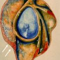
Standard radiographs usually add little information to the evaluation of the throwing athlete with chronic anterior laxity, in comparison with instability from an acute traumatic episode because a humeral head impaction fracture or glenoid rim fracture is less likely. MR arthrography is the modality of choice for assessing glenohumeral instability and diagnosing labroligamentous injuries ( Fig. 2 ).
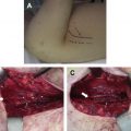
Treatment is aimed at relieving pain, restoring motion, and increasing strength to improve the dynamic stability of the shoulder, with most throwing athletes responding well to an appropriate nonoperative protocol. Because the capsulolabral complex has no contractile elements, there is no effective noninvasive means to restore tension in these structures. However, capsular stretch can often be compensated for by improving dynamic stabilization while avoiding positions of abduction and external rotation. When a trial of nonoperative management fails to relieve symptoms, surgical intervention is often warranted. Diagnostic arthroscopy can reveal anteroinferior capsular lesions, anterior labral damage, humeral head subluxation, or undersurface tears of the supraspinatus or infraspinatus tendons. The drive-through sign is positive when the arthroscope can be easily passed between the humeral head and anterior glenoid with the arm in external rotation, resulting from a lax anterior capsule. With gross laxity of the capsular or ligamentous structures, a stabilization procedure may be indicated, to reinforce the anterior capsule. Anterior capsulolabral reconstruction, as described by Altchek and colleagues, involves making a T-shaped incision in the anterior capsule, with advancement of the inferior flap superiorly and the superior flap medially to reduce capsular laxity both anteriorly and inferiorly. Anterior capsulorrhaphy can also be accomplished arthroscopically with the use of sutures and suture anchors. The treating surgeon must be cognizant to not overtighten the anterior capsule, resulting in a restricted range of motion. In current orthopedic practice, arthroscopic stabilization procedures are much more commonly performed than open procedures in throwing athletes because they are associated with better outcomes in this population.
SLAP Lesions
SLAP lesions can present in the throwing athlete as a dead arm or as inability of the athlete to throw at the preinjury velocity because of pain and subjective feelings of discomfort in the shoulder. It is postulated that SLAP tears can occur as a result of the peel-back mechanism with the arm in the cocked position of abduction and external rotation. In late cocking, the vector of the biceps tendon shifts posteriorly, which transmits a torsional force to the posterior superior labrum. If the superior labrum is not well fixed to the glenoid, this force will cause the labrum to rotate medially over the glenoid rim onto the posterosuperior scapular neck. In throwing athletes, SLAP lesions may be attributed to repetitive traction from overuse.
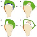
Four types of SLAP lesions are described ( Fig. 3 ). Type I lesions involve fraying and degeneration of the superior labrum, with the labrum and biceps anchor remaining fixed to the glenoid rim. Type II lesions include detachment of the superior labrum and biceps anchor from their insertion. In Type III lesions, a bucket-handle tear occurs in the superior labrum, while the periphery of the superior labrum and biceps anchor remain attached to the glenoid rim. In Type IV tears the bucket-handle tear extends into the biceps anchor. Type II SLAP tears can be subdivided based on anatomic location of the tear: anterior to the biceps insertion, posterior, and combined anterior and posterior. In a study by Burkhart and coworkers, 44 pitchers with Type II SLAP lesions were all found to have tight posteroinferior capsules evident on physical examination as a marked decrease in internal rotation with the arm in 90° abduction. It is postulated that, as the arm abducts and externally rotates during late cocking, a tight posteroinferior capsule pushes the humeral head superiorly, causing a posterosuperior shift in the glenohumeral fulcrum. The end result is an increased force at the biceps-labral attachment, further contributing to the peel-back mechanism.
The throwing athlete with an SLAP lesion often reports sharp pain in the shoulder during late cocking, as the arm is abducted and brought into a position of maximal external rotation. On physical examination, an audible popping or snapping is often heard with shoulder motion. In addition, dynamic scapular winging can often be observed, caused by periscapular muscle weakness and a tight posteroinferior capsule. Various tests can help the examiner determine the location of the SLAP lesion, including the Speed test, O’Brien cross-arm test, and Jobe relocation test.
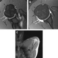
Radiographic evaluation is usually normal in patients with isolated SLAP tears. If an SLAP tear is suspected on clinical examination, the orthopedist usually turns to MR imaging, although MR arthrography is gaining more widespread use because of reported increased sensitivity ( Fig. 4 ). Specific questions the shoulder surgeon would want to know from MR imaging have been presented by Jost and Gerber, including the following: is the biceps insertion normal; is there an SLAP lesion; what type of SLAP; and is there tendinopathy (of the biceps tendon). However, it is essential to examine the entire shoulder region because associated injuries are common with SLAP tears. With MR arthrography, Bencardino and colleagues found that SLAP lesions were associated with partial rotator cuff tears in 42% of patients, frayed or lax inferior glenohumeral ligaments in 26%, Bankart lesions in 16%, Hill-Sachs lesions in 16%, chondral lesions in 16%, loose bodies in 10%, complete rotator cuff tears in 5%, and posterior labral tears in 5%.
Related posts:
 Magnetic Resonance Imaging of Rotator Cuff Disease and External Impingement
 Internal Impingement Syndromes
 The Rotator Cable: Magnetic Resonance Evaluation and Clinical Correlation
Magnetic Resonance Imaging of the Pediatric Shoulder
The Rotator Cable: Magnetic Resonance Evaluation and Clinical Correlation
Magnetic Resonance Imaging of the Pediatric Shoulder
 Entrapment Neuropathies of the Shoulder
 Anatomic Variants and Pitfalls of the Labrum, Glenoid Cartilage, and Glenohumeral Ligaments
Stay updated, free articles. Join our Telegram channel

Full access? Get Clinical Tree


