Chapter 24 Thoracic Zygapophysial Joint Nerve (Medial Branch) Injection, Posterior Approach
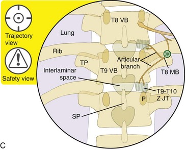
Thoracic medial branch nerve injections are indicated for the diagnosis of axial thoracic (i.e., mid back) pain that typically originates from zygapophysial (i.e., facet) joint sprains, contusions, or osteoarthritis. These injections are performed with the use of a posterior approach. The needle tip is placed at the superior lateral edge of the transverse process, where each medial branch nerve is located as it travels around the intertransverse ligament before it continues medially toward the cephalad neuroforamina, where it then joins the somatic nerve. As a result, the medial branch nerves are not labeled for the transverse process that they cross but rather for their originating somatic nerves. For example, the T8 medial branch crosses the superior lateral edge of the T9 transverse process as shown in Figure 24–1, C. The T1 through T10 medial branch nerves are injected in this manner, whereas the T11 and T12 medial branch nerves are injected with the use of the same landmarks that are used when injecting the lumbar medial branch nerves.
Each thoracic zygapophysial joint receives innervation from two medial branch nerves: from the exiting somatic nerve at the level of the thoracic zygapophysial joint and from the one above it (see Figure 24–6). For example, the T8-T9 zygapophysial joint receives innervation from the T7 and T8 medial branch nerves.
Note: Please see page ii for a list of anatomical terms/abbreviations used in this book.
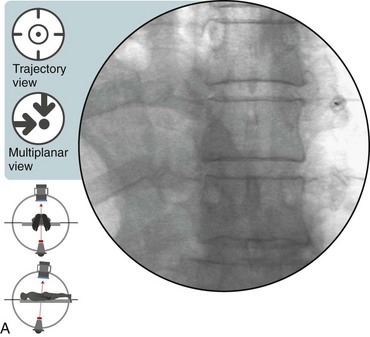
 Trajectory View (Figure 24–1)
![]() Notes on Positioning in the Trajectory View
Notes on Positioning in the Trajectory View
 With this approach, the needle tip is posterior to the superior lateral edge of the transverse process over which the targeted medial branch nerve passes. The needle is not “walked off” of the edge of the transverse process. Rather, the needle is advanced safely by alternating between AP and lateral fluoroscopic views until the tip contacts the superior lateral edge of the transverse process.
 A different spinal needle can be used as a marker to identify the injection level as demonstrated in Chapter 22.
Stay updated, free articles. Join our Telegram channel

Full access? Get Clinical Tree


