Thorax
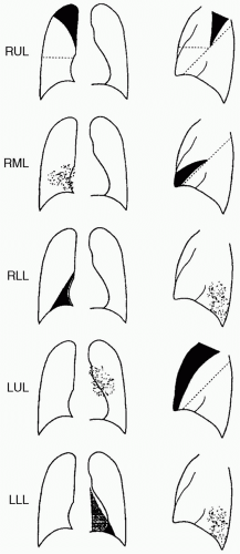 Figure 69-1. Patterns of lobar atelectasis. Roentgenographic Signs of Atelectasis |
Direct
Displacement of interlobar fissures — the only direct sign of atelectasis.
Indirect
Focal increase in density.
Hemidiaphragm elevation — more prominent in lower lobe atelectasis than in upper lobe atelectasis.
Tracheal shift — occurs only with upper lobe atelectasis.
Cardiac shift — occurs variably with lower lobe atelectasis.
Hilar displacement — more prominent in upper lobe atelectasis than in lower lobe atelectasis.
Absence of air bronchogram — only if resorptive type atelectasis.
Nonvisualization of interlobar artery — differentiates lower lobe atelectasis from pleural effusion.