Thorax, Mediastinum, Heart, and Great Vessels: Thymus Size from 0 to 2 Years of Age


10.1055/b-0034-87953

Thorax, Mediastinum, Heart, and Great Vessels: Thymus Size from 0 to 2 Years of Age


Material and Methods


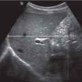
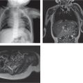
Mediastinal ultrasonography was performed in 151 infants (79 boys and 72 girls; Table 6.2 ). All children were healthy and had no stress factors affecting their thymic size. The maximum transverse diameter, right lobe AP, and left lobe AP were assessed. Perpendicular to the transverse plane, the longest craniocaudal dimension (length) was assessed. The thymic index was calculated by multiplying the transverse diameter by the largest sagittal area ( Fig. 6.3 ).


Table 6.2 Thymus size from 0 to 2 years of age


Thymic dimensions (cm)


Age (months)


Number of cases


Transverse (mm)


AP right (mm)


AP left (mm)


Length (mm)


Thymic index (cm)


Premature


20


2.9. (0.3)*


1.5 (3.7)


1.5 (0.3)


3.1 (0.4)


11.9 (3.9)


0–1


25


3.3 (0.3)


1.6 (0.3)


1.8 (0.3)


3.6 (0.3)


18.1 (6.7)


1–2


21


3.6 (0.6)


1.9 (0.3)


2.0 (0.3)


3.9 (0.4)


25.4 (9.4)


2–3


11


3.7 (0.5)


1.9 (0.3)


2.1 (0.3)


3.8 (0.3)


22.3 (6.9)


3–4


15


3.8 (0.4)


1.9 (0.5)


2.2 (0.6)


4.0 (0.4)


26.8 (10.3)


4–5


7


4.0 (0.6)


2.0 (0.6)


2.2 (0.4)


4.1 (0.3)


29.7 (17.6)


5–6


8


3.9 (0.2)


1.9 (0.4)


2.3 (0.3)


4.0 (0.2)


24.2 (9.3)


6–8


8


3.6 (0.5)


1.8 (0.4)


1.9 (0.5)


3.6 (0.6)


22.2 (8.9)


8–10


14


3.7 (0.5)


1.7 (0.5)


1.9 (0.5)


3.7 (0.4)


21.5 (6.8)


10–12


5


3.7 (0.5)


1.9 (0.6)


1.8 (0.5)


3.6 (0.2)


23.2 (7.2)


12–18


7


2.8 (0.5)


1.2 (0.5)


1.4 (0.4)


3.2 (0.9)


17.2 (6.4)


18–24


10


3.3 (0.4)


1.2 (0.4)


1.2 (0.3)


3.4 (0.7)


15.4 (5.6)


*Standard deviation.


Reproduced with permission of American Institute of Ultrasound in Medicine – AIUM from Yekeler E, Tambag A, Tunaci A, et al. Analysis of the thymus in 151 healthy infants from 0 to 2 years of age. J Ultrasound Med 2004;23:1321–1326; permission conveyed through Copyright Clearance Center, Inc.

Fig. 6.3a, b Thymus. Maximal transverse diameter (a) and largest sagittal area (b).

Only gold members can continue reading. Log In or Register to continue

Stay updated, free articles. Join our Telegram channel

Jul 12, 2020 | Posted by in PEDIATRIC IMAGING | Comments Off on Thorax, Mediastinum, Heart, and Great Vessels: Thymus Size from 0 to 2 Years of Age

Full access? Get Clinical Tree

Get Clinical Tree app for offline access