Thorax: The Lungs


10.1055/b-0034-87851

Thorax: The Lungs


Diffuse Lung Disease


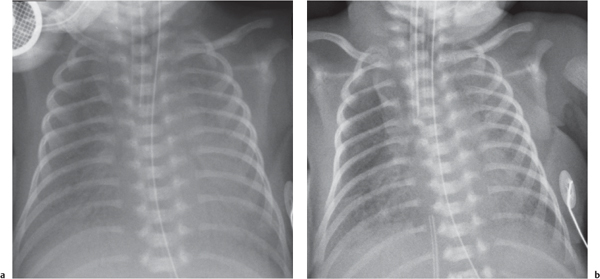
Lung Disease in the Neonate


The differential diagnosis for diffuse lung disease in the neonatal period is relatively narrow. The final diagnosis is reached by a combination of clinical and radiologic findings. Knowing the gestational age of the neonate is essential to constructing a sensible list of possibilities.


Table 1.1 Lung disease in the neonate

Diagnosis


Findings


Comments


Hyaline membrane disease (HMD)/surfactant deficient disease/respiratory distress syndrome


Fig. 1.1a, b


On initial radiograph: Diffuse hypoaeration with small lung volumes, diffuse reticulogranular opacification with or without air bronchograms. Opacification may clear patchily following surfactant administration.


Occurs predominantly in neonates under 36–40-wk gestation. Appearances are due to diffuse alveolar microatelectasis owing to surfactant deficiency. This classic appearance is rare today owing to improved perinatal care, particularly the use of maternal glucocorticoids and direct tracheal instillation of exogeneous surfactant.


Transient tachypnea of the newborn


Fig. 1.2


Normal or mildly increased lung volumes. Diffuse mild predominantly reticular opacification. Small pleural effusions common. May simulate HMD, meconium aspiration syndrome, or neonatal pneumonia, but unlike these clears rapidly (1–2 d).


Associated with cesarean delivery, rapid labor, and low birth weight.


Meconium aspiration syndrome (MAS)


Fig. 1.3


Lung overinflation with radiating perihilar coarse opacities (“ropelike”).


Radiologic features result from small airway obstruction and inflammation, resulting in alternating areas of overinflation and atelectasis. Occurs in term and postterm neonates following fetal distress. Highly susceptible to pneumothorax. Overall mortality up to 25%.


Neonatal pneumonia


Variable patterns. May simulate transient tachypnea of the newborn (TTN) or HMD. Pleural effusion present in two-thirds.


Group B Streptococcus most common agent. Chlamydia pneumoniae usually presents later (typically around 6 wk)


Neonatal heart failure


Fig. 1.8, p. 8


May simulate TTN or HMD. Cardiomegaly may be present, but not usually in cases of abnormal pulmonary venous drainage (e.g., total anomalous pulmonary venous return [TAPVR]).


Common causes presenting in first week of life include left ventricular outflow obstruction, coarctation, aortic stenosis, hypoplastic left heart. Obstructed pulmonary venous return: TAPVR, stenosis of common pulmonary vein, mitral stenosis, cor triatrium.


Myocardial disorders: myocardial ischemia, myocarditis, dysrhythmia.


High output states: vein of Galen malformation, hepatic hemangioendothelioma.


Pulmonary lymphangiectasia


Fig. 1.4a, b, p. 6


Coarse interstitial infiltrate. Occasional septal lines. Peribronchial thickening may result in air trapping. Large chylous effusion often present.


Rare. Indistinguishable from lymphangiomatosis radiologically.

Fig. 1.1a, b Hyaline membrane disease. (a) Chest radiograph in neonate born at 28 weeks’ gestation with respiratory distress. The lungs are of small volume despite ventilation, with a diffuse infiltrate. (b) Chest radiograph taken 48 hours later in the same child following endotracheal surfactant administration. There has been patchy clearing of the diffuse infiltrate.
Fig. 1.2 Transient tachypnea of the newborn. Chest radiograph in a 38-week-gestation neonate delivered by emergency cesarean section. There is a diffuse fine interstitial infiltrate. The lung volumes are marginally increased with seven anterior ribs visible above the diaphragm. The child required overnight nasal positive pressure support only.
Fig. 1.3 Meconium aspiration syndrome. Chest radiograph in term neonate following emergency cesarean section. The lungs are overinflated with coarse opacities throughout the lungs. Note malposition of a nasogastric (NG) tube (black arrow) and umbilical venous catheter (white arrow).
Fig. 1.4a, b Pulmonary lymphangiectasia. (a) Chest radiograph in a neonate with a large right-sided pleural effusion at birth, now drained. There is diffuse lung reticulation with numerous interstitial lines (white arrow). (b) High-resolution CT (HRCT) in same child shows numerous linear opacities representing thickened interlobular septa (arrows). Biopsy confirmed pulmonary lymphangiectasia.


Diffuse Lung Disease Beyond the Neonatal Period


Characterizing diffuse lung disease requires a determination of the characteristics of the opacification, as discussed subsequently.


Bilateral Homogeneous Opacification: Bilateral Lung “White Out”


  • Increased vascular opacities


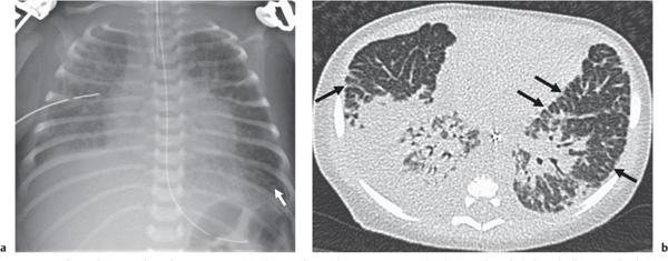
  • Diffuse airspace opacification


  • Diffuse peribronchial opacification


  • Reticulonodular opacification


  • Cystic lung disease


  • Nodular opacification: miliary pattern


  • Generalized patchy opacification


  • Diffuse hypertransradiancy/lung overinflation


Clearly, there is overlap between these groups.


Table 1.2 Bilateral homogeneous opacification: bilateral lung “white out”

Diagnosis


Findings


Comments


Deep expiration


Fig. 1.122, p. 72


All contours obliterated. No air bronchograms. Degree of opacification does not relate to clinical status of child. Trachea often buckled, convex to the right (left if right aortic arch). Opacification may appear almost complete.


Exposure occurs at end of cry. Requires repeat film in inspiration.


Pulmonary hypoplasia


Small, opaque lungs may be present initially. Ribs may be short or downward sloping. Thorax may be bell-shaped. Evidence of secondary cause may be present (e.g., skeletal dysplasia).


Usually secondary to external thoracic compression of lungs in utero: mass (e.g., diaphragmatic hernia, large congenital cystic adenomatoid malformation [CCAM]), oligohydramnios usually due to renal failure [i.e., Potter sequence]) or rib cage abnormality (e.g., Jeune syndrome or in major abdominal wall defect). Idiopathic (primary) form rare, with frequent associated anomalies.


HMD (also known as respiratory distress syndrome [RDS], surfactant deficient disease)


Fig. 1.1, p. 5


More commonly with reticulogranular pattern progressing to air space, but initial opacification may be homogeneous due to diffuse microatelectasis.


Occurs predominantly in neonates under 36–40-wk gestation. Appearances are due to diffuse alveolar microatelectasis owing to surfactant deficiency. This classic appearance is rare today owing to improved perinatal care, particularly the use of maternal glucocorticoids and exogeneous surfactant.


Bilateral large pleural effusions


Diffuse white out appearance on supine film.


Underventilated lungs following intubation


Fig. 1.5


Endotracheal tube (ETT) present: may be abnormally sited (e.g., in esophagus or bevel abutting tracheal wall).


This may be due to endotracheal tube obstruction or be intentional (e.g., when treating meconium aspiration with extracorporeal membrane oxygenation [ECMO]).


Severe diffuse pulmonary hemorrhage


Lung volumes often preserved.


Causes include bleeding diathesis, vasculitis, persistent pulmonary hypertension of the newborn. Occasionally following surfactant therapy.


Severe pulmonary edema


Cardiac enlargement often present.


Fig. 1.5 Intentional hypoventilation. Chest radiograph of a neonate with severe meconium aspiration undergoing venovenous ECMO: the single ECMO cannula lies in the region of the right atrium (arrow). The lungs are completely opacified as a result of minimized ventilation parameters to protect them from barotrauma and rest the lungs for repair.


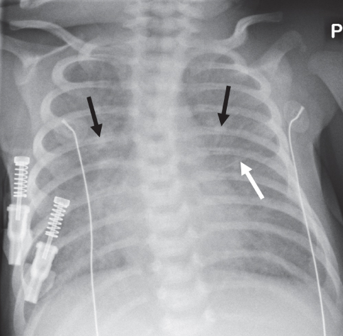
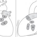
Increased Vascular Opacities

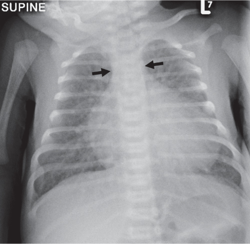
Distinction between pulmonary venous and arterial dilatation is not always straightforward, and the two may coexist. In venous dilatation, the enlarged vessels are less well defined than in arterial dilatation and have a vertical course in the upper zones with a horizontal course in the lower zones. In pulmonary arterial dilatation (pulmonary plethora), dilated arteries radiate from the hilum: the central pulmonary arteries and pulmonary outflow tract may also be dilated.


Table 1.3 Pulmonary venous dilatation

Diagnosis


Findings


Comments


Left ventricular outflow limitation


Cardiomegaly usually present.


Aortic coarctation, particularly in neonatal presentation, congenital aortic stenosis, hypoplastic left heart syndrome.


Left ventricular dysfunction


Fig. 1.32, p. 24


Cardiomegaly usually present.


Myocarditis, anomalous coronary circulation, dilated cardiomyopathy.


Obstructed venous return


Fig. 1.6


Heart size often normal.


Most commonly infracardiac total anomalous pulmonary venous drainage (TAPVD), also mitral valve disease.


High-output cardiac failure


Heart size may be normal or increased.


Consider if echocardiogram shows good left ventricle. Causes include vein of Galen malformation and hemangioendothelioma. Severe anemia in older child.

Fig. 1.6 Pulmonary venous hypertension. Chest radiograph in a neonate with obstructed infracardiac TAPVD. There are ill-defined enlarged central vessels (black arrows) with evidence of interstitial and alveolar pulmonary oedema (white arrow).


Table 1.4 Pulmonary arterial dilatation

Diagnosis


Findings


Comments


Left-to-right shunts


Fig. 1.7


Fig. 1.153, p. 87


Right atrial and ventricular enlargement in ASD, biatrial, and right ventricular enlargement in ventricular septal defect (VSD). Often just mild cardiomegaly and large pulmonary artery segment.


ASD, VSD, PDA, and atrioventricular septal defect (AVSD)/endocardial cushion defect most common lesions.


Admixture lesions


Fig. 1.8


Pulmonary plethora in cyanotic child.


Causes include transposition of great arteries; truncus arteriosus; TAPVD; tricuspid atresia with VSD; double outlet right ventricle; single ventricle.


Pulmonary disease


Severe pulmonary disease resulting in pulmonary hypertension.


Cystic fibrosis (CF) most common cause in children. Also bronchiolitis obliterans.


Pulmonary hypertension: other causes


Fig. 1.16, p. 14


Lungs often normal in appearance. May show dilated central arteries and “pruned” peripheral vasculature (see section on hilar enlargement).


Including primary pulmonary hypertension, pulmonary veno-occlusive disease, recurrent pulmonary embolism, partial anomalous pulmonary venous return.


Poststenotic


Fig. 1.159, p. 90


Usually just pulmonary trunk ± proximal left pulmonary artery visibly dilated.


Fig. 1.7 Pulmonary overcirculation. Chest radiograph in an infant with a large atrioventricular septal defect, demonstrating marked pulmonary plethora with superadded pulmonary edema due to “overcirculation.” Note the fissural fluid (arrow).
Fig. 1.8 Pulmonary plethora. Chest radiograph in an infant with dextro-transposition of the great arteries. There are well-defined enlarged vessels throughout the lungs. Note the narrow superior mediastinum (between arrows) due to superimposition of the aorta and main pulmonary artery, producing the “egg on a string” appearance.


Table 1.5 Mixed

Diagnosis


Findings


Comments


Left-to-right shunt with heart failure


Fig. 1.8


Large heart with large pulmonary vessels and diffuse air-space/interstitial shadowing.


Heart failure may develop due to pulmonary overcirculation. Common with large septal defects, AVSD. May require pulmonary artery banding prior to definitive treatment.


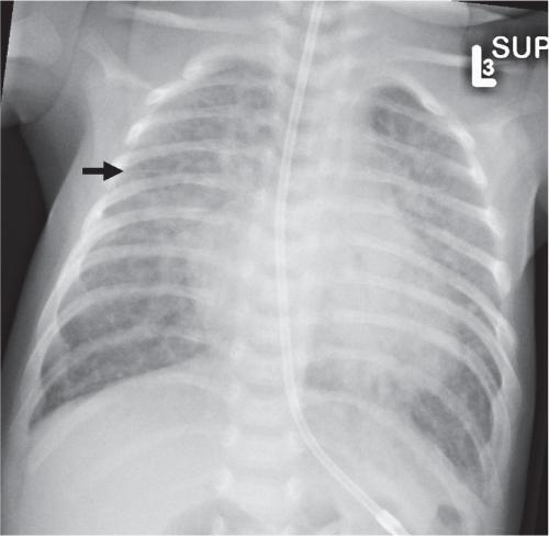
Diffuse Airspace Opacification


Table 1.6 Diffuse air-space opacification

Diagnosis


Findings


Comments


Cardiogenic pulmonary edema


Fig. 1.8


Heart usually but not always enlarged.


In the neonatal period, cardiogenic pulmonary edema is most commonly due to left-to-right shunts with pulmonary overcirculation. In later life, more common causes include myocarditis and dilated cardiomyopathy.


Acute (“adult”-type) respiratory distress syndrome (ARDS)


Diffuse bilateral air-space opacification within 24–48 h of precipitating event. Frequently associated with air leak phenomena in children (pneumomediastinum, pneumothorax, interstitial emphysema).


Common precipitating events are septicemia, neurologic disease, and near drowning.


Diffuse pulmonary hemorrhage


Fig. 1.9


Usually normal lung volumes. May be diffuse or patchy air-space shadowing.


Causes include bleeding diathesis, vasculitis including Wegener granulomatosis, following surfactant therapy in neonates and idiopathic (acute idiopathic pulmonary hemosiderosis).


Near drowning


Fig. 1.10


Appearance as ARDS.


May be considered a form of ARDS. Air-space shadowing may reflect aspiration of water resulting in permeability edema (“wet drowning”) or negative pressure edema due to prolonged laryngospasm (“dry drowning”). Infective pneumonia may complicate.


HMD


Fig. 1.1, p. 5


Low lung volumes due to microatelectasis, reticulogranular opacities: air bronchograms may be present.


In premature neonates (see previous sections).

Fig. 1.9 Pulmonary hemorrhage. Chest radiograph in a 13-year-old girl requiring ventilation for atypical pneumonia who developed frank hemoptysis, with hemosiderin-laden macrophages on bronchoalveolar lavage. There is widespread, but patchy air-space shadowing (arrows) that is more extensive on the left.
Fig. 1.10 Pulmonary edema in near drowning. Chest radiograph in a 2-year-old child found unconscious in a pond. There is bilateral central air-space shadowing (arrows).


Diffuse Peribronchial Opacification

Peribronchial opacification is a common finding in pediatric chest radiology, particularly in younger children. There appears to be a greater propensity for respiratory infections to involve small- and medium-sized airways in younger children. Smaller airway luminal size means peribronchial thickening often results in airway obstruction, usually manifest as air trapping.


Table 1.7 Diffuse peribronchial opacification

Diagnosis


Findings


Comments


Viral pneumonia/bronchiolitis


Fig. 1.11


Peribronchial thickening particularly in hilar regions. Airway obstruction results in varying degrees of diffuse air trapping, patchy air trapping, and areas of atelectasis.


The distinctions between viral bronchitis, bronchiolitis, and bronchopneumonia are arbitrary.


Common viral agents include respiratory syncytial virus (RSV), influenza, parainfluenza, adenoviruses, and enteroviruses.


Recent evidence suggests that age is a better predictor of radiographic pattern than infecting organism: peribronchial opacities and lung hyperaeration are more common in infants, and alveolar opacities are more common in the older child.


Mycoplasma pneumonia


May mimic viral pneumonitis. The classic pattern is segmental or lobar interstitial changes (relatively specific feature) progressing to air-space shadowing. Hilar nodal enlargement in minority.


Common in school-aged children, accounts for up to 30% of childhood pneumonias.


Bronchial asthma


Seventy-five percent of radiographs in acute asthma will demonstrate peribronchial thickening and hyperaeration. Focal infiltrates and atelectasis in 25%. In chronic asthma, similar changes may be present in approximately 20%.


Be aware of asthma mimics. Conditions commonly mislabelled as asthma in children include CF, constrictive obliterative bronchiolitis, chronic foreign body aspiration, vascular ring, mediastinal mass, and tracheal mass lesion.


CF


Fig. 1.12a, b


Milder cases may demonstrate peribronchial thickening as only abnormality and may simulate other conditions causing peribronchial thickening.


Should be considered in any child with recurrent respiratory problems.


Bronchiectasis


Fig. 1.12a, b


Fig. 1.31, p. 24


CXR: tram-track and ring shadows in areas of involvement on plain radiograph, with bronchial dilatation (airway larger than accompanying pulmonary artery).


HRCT: for definitive diagnosis.


CF most common cause. Postinfectious causes now rare since pertussis and measles vaccination introduced. Primary immunodeficiencies, primary ciliary dyskinesia, allergic bronchopulmonary aspergillosis, amongst other causes.


Pulmonary venous hypertension


Fig. 1.6, p. 7


Fig. 1.7, p. 8


May simulate viral infection. Large heart may be present. Septal lines suggestive but rare. Small effusions, larger on the right, often present.


In neonate and infants, frequently due to large left-to-right shunts. In older children, consider myocarditis and dilated cardiomyopathy.


TTN


Fig. 1.2, p. 5


Normal or mildly increased lung volumes. Diffuse mild predominantly reticular opacification. Small pleural effusions common. May simulate HMD, MAS, or neonatal pneumonia, but unlike these clears rapidly (1–2 d).


In neonates only.

Fig. 1.11 RSV bronchiolitis. Chest radiograph of a 4-week-old girl with RSV-positive bronchiolitis. The lungs are overinflated. There is a diffuse peribronchial infiltrate (white arrows), with upward bowing of the horizontal fissure indicating early right upper lobe (RUL) collapse (black arrow)
Fig. 1.12a, b Cystic fibrosis. (a) Chest radiograph in a 10-year-old girl with known CF. There is lung overinflation with diaphragmatic flattening. There is some mild central bronchial wall thickening (white arrow) and some subtle ring and tram-track opacities in the basal regions (black arrows). (b) HRCT in the same patient. There is diffuse mild bronchiectasis (white arrows). There are some subtle centrilobular nodularities (black arrow) representing areas of small airways mucus plugging.


Reticulonodular Opacification

A reticulonodular pattern (i.e., one consisting of discrete nodular and linear opacities) is typically due to disease of the pulmonary interstitium. The exact pattern depends on the distribution of changes. Thickening of the peribronchovascular interstitium results in peribronchial thickening centrally, but peripherally results in branching nodular opacities. Thickening of the interlobular septa results in septal lines: these may appear as vertically oriented lines in the upper zones, horizontally oriented lines in the lung periphery, or diffuse spidery lines throughout the lungs. Lung reticulation on a radiograph may also result from cystic change due to overlapping cyst walls, as in diffuse cystic lung diseases, from end-stage fibrotic changes (“honeycombing”), and from overlapping bronchial walls in severe bronchiectasis.


Table 1.8 Reticulonodular opacification

Diagnosis


Findings


Comments


HMD


(see section on diffuse neonatal lung disease)


Neonatal pneumonia


TTN


Pulmonary lymphangiectasia


Fig. 1.4, p. 6


Interstitial pattern often with septal lines. Pleural effusions very common and often large.


Primary pulmonary lymphangiectasia usually presents in neonatal period and is frequently fatal. May be secondary to congenital heart disease, particularly anomalous pulmonary venous connection, or following lymphatic injury, usually surgical. Also associated with Turner and Noonan syndromes.


Viral pneumonitis


Fig. 1.11, p. 11


Peribronchial thickening the norm.


Mycoplasma pneumonia


Patchy or unifocal interstitial infiltrate, often with hilar adenopathy.


Pulmonary venous hypertension


Fig. 1.6, p. 7


Transient appearance of interstitial pulmonary edema, with or without septal lines, often coexisting with central alveolar opacities.


Bronchopulmonary dysplasia (BPD)


Fig. 1.13


CXR: The classic form appears as “bubbly lungs” with an irregular pseudocystic appearance. In the “new” form, the chest radiograph may reveal only diffuse ground-glass opacity, a more uniform interstitial pattern, or a “bubbly” pattern with smaller, more uniformly sized bubbles.


HRCT: Classic features include triangular subpleural and septal thickening and fibrosis, with patchy areas of air trapping. Changes may be more diffuse in “new” form.


A complication of prematurity. Two forms described: classic BPD occurs in children with prolonged positive pressure ventilation ± high inspired oxygen concentrations. “New” BPD probably reflects use of lower pressure ventilation strategies and surfactant therapy, and more reflects pulmonary immaturity in very-low-birth-weight infants rather than effects of barotrauma and oxygen toxicity. The previously described Mikity Wilson syndrome, whereby BPD-like changes developed in nonventilated premature infants, is probably the same condition as new-type BPD.


Langerhans cell histiocytosis (LCH)


Fig. 1.14


Fig. 1.27, p. 21


Nodular opacities ranging from 1 to 10 mm. Cystic changes may reflect pneumatocele formation due to bronchiolar involvement, or cystic degeneration of nodules. Pneumothorax common.


Lung involvement present in approximately 10% at presentation, and up to half with multiorgan involvement.


Interstitial lung disease: other


(see section on HRCT of interstitial lung disease)


Leukemia


May be air-space or interstitial, diffuse, or localized.


Particularly with acute monocytic leukemia. May be first manifestation and may worsen with induction chemotherapy. Changes at least partly due to pulmonary hemorrhage.


Pulmonary fibrosis


Fig. 1.15


“Honeycomb” pattern with volume loss.


The end stage of a variety of interstitial processes.


Pulmonary veno-occlusive disease


Fig. 1.16a, b, p. 14


Pulmonary arterial dilatation with smooth inter-lobular septal thickening, small pleural effusions, and patchy ground-glass attenuation on CT. Hilar adenopathy may also be present. Combination of pulmonary arterial dilatation and smooth septal thickening is highly specific.


A postcapillary counterpart to primary pulmonary hypertension. Very poor prognosis.

Fig. 1.13 Bronchopulmonary dysplasia. Chest radiograph in a 3-month-old infant born at 24 weeks’ gestation, requiring prolonged ventilation. There is a diffuse “bubbly-lung” appearance due to summation of coarse interstitial opacities, and generalized lung overinflation with left basal hypertransradiancy (white arrow).
Fig. 1.14 Langerhans cell histiocytosis. Chest radiograph in a 3-month-old boy with lethargy and a lytic lesion in the left femur. There is a diffuse coarse reticulonodular infiltrate with one or two subtle areas of cavitation (arrow). (See also Fig. 1.27 , a CT of the same child.)
Fig. 1.15 Apical lung fibrosis due to drug reaction. Chest radiograph in an 8-year-old child who underwent conditioning with busulphan 5 years previously for BMT as treatment of relapsed acute myeloid leukaemia. There is apical lung reticulation (arrows) with volume loss evidenced by bilateral hilar elevation.
Fig. 1.16a, b Pulmonary veno-occlusive disease. (a) Chest radio-graph in a 12-year-old girl with progressive breathlessness, weight loss, and pulmonary hypertension. The central pulmonary arteries are enlarged (white arrows). There is extensive linear opacification with septal lines (black arrow). (b) Coronally reformatted CT image in the same patient demonstrates numerous thickened interlobular septa (white arrows). The diagnosis of pulmonary veno-occlusive disease was confirmed at postmortem examination.


Cystic Lung Disease

This category covers the radiographic appearance of diffuse rounded lucencies in the lungs: these may represent cysts or features that merely simulate a cystic appearance (“pseudocystic”; see also the section on focal lung lucencies).


Table 1.9 True cystic diseases

Diagnosis


Findings


Comments


LCH


Fig. 1.27, p. 21


Nodules usually also present. Cysts reflect cavitation of nodules or pneumatocele formation.


Lung involvement present in approximately 10% at presentation, and up to half with multiorgan involvement.


Tuberous sclerosis


Multiple thin-walled cysts, usually small. Interstitial fibrosis in established cases. Chylothorax common.


Indistinguishable from lymphangioleiomyomatosis, with very similar pathogenesis of smooth muscle proliferation in bronchiolar walls.


Lymphangioleiomyomatosis


Thin-walled, randomly scattered cysts with normal intervening parenchyma. Associated chylothorax.


Females of childbearing age only. Exceedingly rare in children.


BPD


Fig. 1.13, p. 13


Not true cysts, but areas of profound air trapping mixed with fibrotic bands producing “bubbly lungs.”


In premature infants, usually those who required intubation and oxygen therapy.


Pulmonary interstitial emphysema


Fig. 1.52, p. 36


Tubulocystic lucencies radiating from hilum. Not true cysts but areas of interstitial gas. Usually transient and diffuse, but may be localized, and may be persistent.


Usually occurs in premature neonates, usually in first week of life. Also occurs with increased frequency in hypoplastic lungs or in the presence of interstitial lung disease. Frequently associated with or precedes pnuemothorax or pneumomediastinum. Predicts development of BPD.


Bronchiectasis


Not true cysts, but saccular changes may simulate cysts.


CF most common cause. Postinfectious now rare since pertussis and measles vaccination introduced. Primary immunodeficiencies, primary ciliary dyskinesia, allergic bronchopulmonary aspergillosis, amongst other causes.


Multiple pneumatoceles


Usually localized, but occasionally multifocal, particularly following hydrocarbon ingestion (see section on focal lung lucencies).


Multiple cavitating lesions


(see section on multiple cavitating lesions)


Such as granulomas or metastases.


Nodular Opacification: Miliary Pattern

A miliary pattern refers to diffuse small nodules that are 1 to 2 mm in size, named as such because they resemble millet seeds.


Table 1.10 Miliary patterns

Diagnosis


Findings


Comments


Miliary tuberculosis (TB)


Fig. 1.17a–c


Discrete small nodules or “snowstorm” appearance, often with lymphadenopathy. Distinct appearance from endobronchially disseminated TB, which shows multiple centrilobular, tree-in-bud opacities.


Usually within 6 months of primary infection. Reflects hematogenous dissemination of primary infection. Liver, spleen, and brain may also be involved.


Histoplasmosis


Fig. 1.44, p. 31


May simulate miliary TB. Resolves into multiple punc-tate calcifications after 9–24 mo.


Rare in nonendemic areas.


Metastatic disease


May exactly simulate miliary TB: may also have “snowstorm” appearance.


Usually thyroid carcinoma, most commonly papillary and follicular.


LCH


Fig. 1.14, p. 13


Nodules often variable in size with coexistent cysts.


Lung involvement present in approximately 10% at presentation, and up to half with multiorgan involvement.

Fig. 1.17a–c Disseminated TB. (a) Miliary TB. Chest radiograph in an 8-year-old child with TB meningitis. There are numerous small nodules throughout the lungs. Calcified lymph nodes are present in the left axilla (arrow). (b) Disseminated endobronchial TB with nodal partial obstruction of bronchus intermedius. Chest radiograph in a 6-month-old with prolonged fever and progressive respiratory distress. There is a diffuse, coarse nodular pattern throughout the lungs. The right middle and lower lobes are overinflated (arrows). (c) Disseminated TB with nodal partial obstruction of bronchus intermedius. CT of the same 6-month-old child shows numerous branching centrilobular nodules (“tree-in-bud” pattern, white arrows) with a large subcarinal nodal mass (black arrow) causing narrowing of the bronchus intermedius with air trapping. TB was isolated from gastric washings.


Generalized Patchy Opacification


Table 1.11 Generalized patchy opacification

Diagnosis


Findings


Comments


Bronchopneumonia


Fig. 1.18


Irregularly distributed alveolar opacities. May be bilateral and diffuse or localized.


Unusual with pneumococcus. Occurs in staphylococcal, Haemophilus influenzae, pertussis, mycoplasma, and viral pneumonias.


Aspiration pneumonitis


Fig. 1.23, p. 20


Opacities more commonly right-sided and in dependent lung regions: posterior segments upper lobes, apical, and posterior basal segments of lower lobes.


Aspiration may be from below (i.e., related to reflux) or from above (impaired swallow, tracheoesophageal fistula, meconium aspiration).


Pulmonary vasculitis


Fig. 1.24, p. 21


Patchy or diffuse air-space opacification, occasionally ill-defined nodules. May develop cavitation.


Changes may reflect patchy pulmonary hemorrhage or inflammatory change. Examples include Wegener granulomatosis and Goodpasture syndrome.


Acute/subacute extrinsic allergic alveolitis


CXR: Acute: patchy, often subtle air-space opacities. Subacute: patchy often reticulonodular pattern. HRCT: centrilobular nodules and patchy ground-glass attenuation.


Known exposure to precipitating antigen and presence of serum precipitins to antigen make diagnosis.


LCH


Often with more discrete nodular densities and cyst formation.


Hodgkin disease


Most commonly nodular, extending from mediastinum along peribronchovascular lymphatics. Pneumonic form shows patchy nonsegmental infiltrates. Usually with ipsilateral hilar/mediastinal nodal enlargement.


Non-Hodgkin lymphoma (NHL), where pulmonary involvement often occurs without nodal involvement.


Sarcoidosis


With or without bilateral hilar enlargement and right paratracheal lymphadenopathy. Variable pattern.


Lung involvement very rare in preteenaged children.


Idiopathic pulmonary hemosiderosis


Patchy alveolar opacities acutely during episodes of hemoptysis and pulmonary hemorrhage.


Eventually develops reticular pattern followed in some by features of established fibrosis.


Clinical triad is of iron deficiency anemia, episodic hemoptysis, and patchy infiltrates on CXR.


Löffer syndrome


Rapidly changing “migratory” infiltrates.


An allergic reaction to a variety of insults, classically parasitic infection but also drugs.


Posttransplant lymphoproliferative disorder (PTLD)


Patchy air-space opacification a rarely reported pattern in PTLD. Hard to distinguish from rejection in lung transplant recipient.


Most common after thoracic (heart, lung, heart/lung) transplantation, but may occur with any transplant. Other thoracic manifestations include multiple nodules (most common), solitary pulmonary nodule, and mediastinal adenopathy.

Fig. 1.18 Bronchopneumonia: pertussis. Chest radiograph in a 2-month-old infant with severe respiratory failure requiring venoarterial ECMO, later proven to be due to Bordetella pertussis. There are patchy coarse air-space infiltrates in the right middle lobe (RML), left lower lobe, lingula, and RUL.


Diffuse Hypertransradiancy

This is a relatively common pattern in children and generally implies diffuse air trapping due to valvelike obstruction of medium and small airways.

Fig. 1.19 Ebstein anomaly with pulmonary oligaemia. Chest radiograph in a neonate with cyanosis. The heart is enlarged, particularly the right atrial contour (arrows). The lungs are markedly oligemic with attenuated vasculature throughout. Echocardiography confirmed Ebstein anomaly.


Table 1.12 Diffuse hypertransradiancy

Diagnosis


Findings


Comments


Bronchiolitis/viral lower respiratory tract infection


Fig. 1.11, p. 11


Diffuse peribronchial opacities, large lung volumes with diaphragmatic flattening. There may be areas of subsegmental, segmental, or lobar atelectasis.


Common viral agents include RSV, influenza, parainfluenza, adenoviruses, and enteroviruses (see section on peribronchial opacification).


Bronchial asthma


In majority with acute asthma, minority with chronic steroid-maintained asthma.


Remember asthma mimics: CF, constrictive obliterative bronchiolitis, chronic foreign body aspiration, vascular ring, mediastinal mass, and tracheal mass lesion.


CF


Fig. 1.12, p. 11


Evidence of bronchiectasis, relative volume loss in the upper lobes, and overall lung overinflation due to air trapping from small and large airway disease.


Meconium aspiration syndrome


Fig. 1.3, p. 5


Neonates only.


(see Table 1.1 )


Cardiac failure (cardiac asthma)


Fig. 1.7, p. 8


Peribronchial edema often results in air trapping. Heart may be enlarged.


Pulmonary oligemia


Fig. 1.19


Attenuated vessels, lung volumes usually normal. Cardiac contour may be abnormal.


In right heart congenital heart disease: tetralogy of Fallot, pulmonary atresia, Ebstein anomaly.


BPD


Fig. 1.13, p. 13


Classic BPD: large lung volumes with multiple small, rounded, cystic lucencies, with intervening irregular opacity.


CT findings: reticular opacities, areas of atelectasis, reduced bronchoarterial ratios, areas of hypertransradiancy, triangular subpleural opacities.


Pattern of BPD is changing with more effective treatment of RDS/HMD. “New” pattern BPD shows more diffuse interstitial changes with less pronounced cystic lucencies.


Chronic aspiration pneumonitis


Recurrent, migratory segmental opacities progressing to fibrotic changes. Peribronchial fibrosis may result in air trapping.


Aspiration either from above (unsafe swallow, tracheoesophageal fistula) or below (reflux-related).


Constrictive obliterative bronchiolitis


Fig. 1.20a, b


Fig. 1.68, p. 45


CXR: May be normal. Diffuse overinflation with attenuated vascularity common. May be associated with large airway changes (bronchiectasis). Asymmetric involvement produces a relatively hyperlucent lung: the Swyer-James-Macleod syndrome.


HRCT: Mosaic attenuation that is accentuated on expiratory sections. Centrilobular nodules with tree-in-bud pattern frequent.


Numerous causes: most common are postinfectious, particularly adenovirus and mycoplasma, connective tissue diseases, chronic lung transplant rejection, graft-versus-host disease following bone marrow transplant (BMT), post toxic fume inhalation, and idiopathic.


Extrinsic tracheobronchial compression


Fig. 1.21


(see Table 1.51 )


Two lobe congenital lobar overinflation (CLO)


Fig. 1.22a–c


Usually left upper and right middle lobe.


Up to 5% of CLO involves two lobes.


High ventilation pressures


Fig. 1.21


Child intubated or on continuous positive airway pressure.


Position of diaphragms on radiograph may be used to guide pressure management, particularly with high-frequency oscillator therapy.

Fig. 1.20a, b Constrictive obliterative bronchiolitis. (a) Chest radiograph in a 14-year-old boy with respiratory failure and prior adenovirus infection. There are extensive areas of hypertransradiancy with attenuated vessels in both lungs (arrows). (b) Expiratory CT section in same child demonstrates areas of hypertransradiancy (black arrows) indicating extensive air trapping. There are a few areas of mild bronchial dilatation (white arrow). This child underwent successful lung transplantation. Obliterative changes in bronchioles confirmed at histologic examination of explanted lungs.
Fig. 1.21 Air trapping due to mediastinal mass. Chest radiograph in an 8-year-old boy requiring ventilation for respiratory distress. There is a large anterior mediastinal mass (white arrows). There is profound air trapping with very marked diaphragmatic flattening (black arrows). This improved with prone positioning. Histologic diagnosis was of T-cell type acute lymphoblastic leukemia.
Fig. 1.22a–c Congenital lobar overinflation involving two lobes. (a) Chest radiograph in an 8-week-old boy with progressive respiratory distress. There is overinflation of the right middle and left upper lobes (LULs), with compressive atelectasis of the RUL (white arrow), left lower lobe (black arrow), and right lower lobe (open arrow). (b) Coronally reformatted CT in the same patient showing hypertransradiancy of right middle (white arrow) and LULs (black arrow). The lingula was spared. (c) Ventilation (right) and perfusion (left) scintigrams in same patient showing nonventilated and perfused right middle (black arrows) and LULs (white arrows). The child had an excellent outcome following surgical resection of the overinflated lobes.

Only gold members can continue reading. Log In or Register to continue

Stay updated, free articles. Join our Telegram channel

Jul 12, 2020 | Posted by in PEDIATRIC IMAGING | Comments Off on Thorax: The Lungs

Full access? Get Clinical Tree

Get Clinical Tree app for offline access