CHAPTER 23
Three-Dimensional Fetal Echocardiography
Ultrasound technology has improved significantly over the last few decades. As three-dimensional (3-D) and four-dimensional (4-D) technologies have improved, so has the interest in applying them to fetal echocardiography. Two primary reasons drive this interest. First, the technology is now readily available on most ultrasound systems. Second, standard two-dimensional (2-D) fetal echocardiography requires considerable expertise to perform and interpret correctly. It can also be very time consuming. Major drawbacks to applying 3-D/4-D technologies to the heart have been acquisition time and the need for cardiac gating. Recent improvements in technology allow for near real-time examination.1
Techniques
Spatiotemporal Image Correlation
STIC is an automated volume acquisition technique.2 It is based on a transducer sweep that acquires a large volume set. The volume set is usually acquired over a period of 7.5 to 30 seconds, at a sweep angle of 20 degree to 40 degrees and a frame rate of 150 frames per second.1,3
The ultrasound system then applies mathematical algorithms to process the volume set. Image gating is accomplished by peak systole signals being automatically detected from the underlying images. The B-mode images are then correlated with the heart rate, and a complete heart cycle is displayed. This 4-D loop can be manipulated to display any acquired scanning plane at any point of the cardiac cycle.4 The advantage of STIC technology is that reconstruction takes place immediately. It can also be combined with other applications such as B-flow, color Doppler imaging, power Doppler imaging, and tissue Doppler imaging.
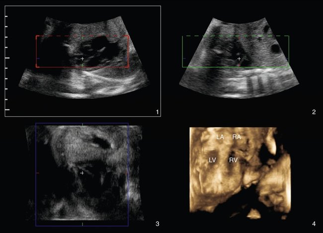
Multiplanar Reconstruction
Multiplanar reconstruction displays the X, Y, and Z axes of an acquired volume set, all on one panel. The operator is then able to manipulate the volume to display any plane within it. The fourth image on the panel will then display a surface rendering of the desired structure or level (Fig. 23–1).1
Spatiotemporal Image Correlation with Tomographic Ultrasound Imaging
TUI-STIC uses STIC technology, which allows the acquisition of cardiac volumes combined with tomographic ultrasound imaging. This allows a variable number (up to nine) of reconstructed 2-D sections to be displayed on a single panel.5,6
Stay updated, free articles. Join our Telegram channel

Full access? Get Clinical Tree


