Fig. 18.1
CT scan with contrast injection shows a “saddle embolus” that straddles the bifurcation of the main pulmonary artery, with massive obstruction of both the right and the left pulmonary arteries
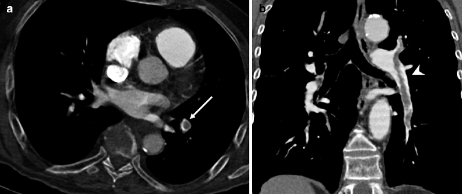
Fig. 18.2
Contrast-enhanced CT axial image at the level of the left atrium (a) and coronal reconstruction (b) in a 67-year-old man with acute pulmonary embolism. When the partial occlusion is observed in a section transverse to the vessel, the “polo mint” sign is seen (arrow). If the artery lies parallel and within the plane of section, the partial occlusion gives rise to the “railway track” sign (arrowhead). An increase in vessel diameter can also be seen
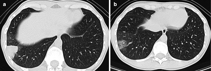
Fig. 18.3
(a) The CT axial section at the lung bases, with parenchymal window, shows a pulmonary infarct in the periphery of the right lung (Hampton hump). (b) One and a half months later, the following CT scan shows the gradual resolution of consolidation, resembling a melting ice cube
Progressive RV failure is the most frequent cause of death related to PE. Increased pulmonary vascular resistance leads to a rise in RV wall tension, resulting in dilation and functional changes. The interventricular septum may protrude into an otherwise normal left ventricle (LV), thus narrowing its lumen and causing incomplete filling, leading to decreased LV output and reduced systemic arterial pressure and coronary artery flow.
Clinical trials have demonstrated that CTPA can play a major role in evaluating the severity of acute PE, as it helps to assess numerous parameters, in particular: RV/LV ratio, interventricular septum bowing, pulmonary artery and superior vena cava dilation, and tricuspidal regurgitation (van der Meer et al. 2005; Collomb et al. 2003; Quiroz et al. 2004; Ghaye et al. 2006). The presence, position, and degree of pulmonary artery occlusion may be quantified by using vascular obstruction indexes. There is considerable debate concerning the use of these data. Despite their proven value in quantifying arterial obstruction, their use is limited by their complexity. Recently, the Qanadli score (Qanadli et al. 2001), based on evaluation of all arterial branches involved in the obstruction, has been replaced by a score proposed by Ghanima et al. (2007), which is easier to apply in clinical practice and has proved to be a valid indicator of prognosis (Attinà et al. 2011).
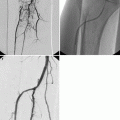
Acute PE can resolve without sequelae, even though in many cases, embolic remnants are still visible at MDCT 6–12 months after the event. Non-resolution of acute emboli, which later undergo fibrosis, leading to mechanical obstruction of PAs and increasing arterial resistance, can lead to chronic thromboembolic pulmonary hypertension (CTEPH) in a small percentage of patients (Hoeper et al. 2006). The distinction between acute versus chronic PE is possible on CTPA because chronic emboli appear as partial and eccentric occlusions that adhere to the arterial wall forming obtuse angles (Fig. 18.4); the chronic thrombus may contain calcifications and may show sign of recanalization, with contrast medium penetrating in its context. Some vessels may be completely occluded (Fig. 18.5) and may show a smaller diameter than adjacent vessels of the same order (clot retraction). In addition, intraluminal plicae, bands, webs, or pouching can be appreciated (Wittram et al. 2005).
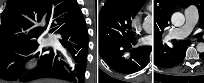
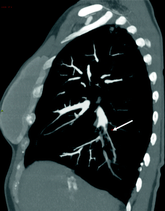
Fig. 18.4
MDTC MIP sagittal reconstruction (a) in a case of chronic pulmonary embolism shows partial occlusions at the level of the right pulmonary artery and lower right lobe (arrows). (b) MDTC axial image at the level of the right lower lobe pulmonary artery (arrow). (c) Axial image shows how chronic emboli adhere to the vessel wall forming obtuse angles (arrow). Dilation of right pulmonary artery can also be appreciated
Fig. 18.5
MDTC MIP sagittal reconstruction in a patient with chronic pulmonary embolism shows complete occlusion and fibrotic retraction of right lower lobe pulmonary artery (arrow) with clot remnants inside
18.1.5 Other Diagnostic Techniques
Pulmonary arteriography is rarely used, especially in elderly patients and in critical conditions, because of a not trivial risk of renal failure or other serious complications. Currently, it is indicated in patients undergoing embolectomy or catheter-based thrombolysis (Stein et al. 1992).
Magnetic resonance angiography is not routinely used because it has the disadvantages of a limited spatial resolution, access problems in the emergency room, and a still restricted availability. Its role is therefore subordinated to MDCT and limited to patients with absolute contraindications to iodinated contrast media (Kanne and Lalani 2004).
Echocardiography allows assessment of RV function but rarely direct visualization of emboli and only those in the main pulmonary arteries. It plays a role of diagnostic support in critically ill patients to demonstrate the hemodynamic changes (dilation or hypokinesis of the RV, dilation of the inferior vena cava with lack of inspiratory collapse, etc.) (Goldhaber 2002).
18.1.6 PE in Acute COPD Exacerbation
The occurrence of PE in patients with COPD is important because of combined morbidity and mortality. In fact, PE may aggravate the already precarious respiratory state of these fragile patients. PE may be a relatively common but unrecognized cause of acute COPD exacerbations, since cough, dyspnea, and chest pain are frequently found in both conditions (Stein et al. 2007).
Patients with COPD have an increased risk of PE, estimated at around twice that of patients without COPD (Sidney et al. 2005). The prevalence of PE in patients with COPD is variable in different studies, probably due to different inclusion criteria (Rizkallah et al. 2009). Two different studies reported PE in about 25 % of patients presenting with COPD exacerbation of noninfectious etiology (Tillie-Leblond et al. 2006; Mispelaere et al. 2002). However, the prevalence of PE is very low in patients admitted in the emergency department for acute COPD exacerbation (3.3 %) (Rutschmann et al. 2007).
While COPD remains a clinical diagnosis, PE requires objective confirmation of clots by an imaging study to establish an appropriate anticoagulation therapy. At chest x-ray, PE cannot be easily distinguished from COPD exacerbation, because RV hypertrophy and pulmonary arteries dilation can lead to confusing results. Also the interpretation of lung scintigraphy can be difficult in patients with COPD, because it might lead to an overestimation of PE. On the contrary, the performance of CT is not altered in patients with COPD (Rutschmann et al. 2007).
In most cases, Doppler ultrasonography of lower limbs may be the appropriate first imaging study if there is concern for a thromboembolic diagnosis particularly when respiratory symptoms are equivocal.
18.1.7 Tumor Embolism
Tumor embolism is a rare complication of cancer in which neoplastic cells from an organ enter the systemic venous and pulmonary arterial system, where they lodge, and obstruct flow, without invading the vessel wall. This condition differs from widespread blood-borne metastases, in which tumor invades the vessel wall and acquires its own blood supply (Fig. 18.6).
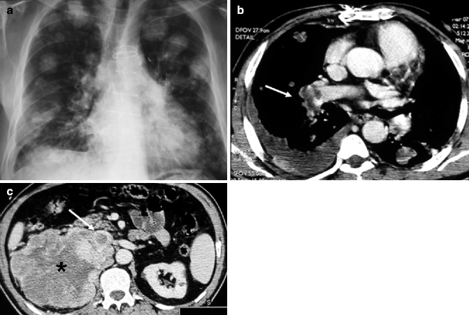
Fig. 18.6
A case of renal cancer with multiple lung metastases. (a) Chest x-ray shows multiple parenchymal opacities of both lungs. (b) MDTC axial image shows a tumor embolus of right pulmonary artery (arrow). Subpleural solid lesions and right pleural effusion can also be appreciated. (c) Right renal large tumor (asterisk) with associated invasion and thrombotic occlusion of inferior vena cava (arrow)
Hemodynamically significant tumor emboli are rare, but the occlusion of many small pulmonary vessels can give rise to severe dyspnea, pleuritic chest pain, and signs of cor pulmonale that develop over weeks to months (Roberts et al. 2003). The mortality of this condition is very high, but it is rarely diagnosed ante mortem and tumor emboli are a frequent finding at autopsy in patients with history of solid tumors (Iwakami et al. 2009; Corradi et al. 2006; Nakamura et al. 2004). The primary tumors more commonly associated with tumor embolism are hepatocarcinoma, lung (Fig. 18.7), breast and renal carcinoma, gastric and prostate cancer, and choriocarcinoma (Chan et al. 1987).
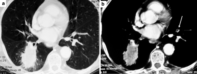
Fig. 18.7
(a) MDTC axial image shows a lung adenocarcinoma in the lower right lobe. (b) The same image with mediastinal window shows a paraneoplastic thrombus in a segmental artery of left lung (arrow)
Chest x-ray is usually normal or can show nonspecific pulmonary shadows, possibly infarcts. Lung scintigraphy characteristically reveals multiple subsegmental mismatched defects. On MDCT, diagnosis can be occasionally suggested by micronodules or diffuse pulmonary infiltrates. An unusual pathologic finding is represented by thrombotic microangiopathy (Yao et al. 2001) in which tumor microemboli cause fibrocellular proliferation of the intima with resulting thrombosis and luminal obliteration with subsequent severe pulmonary hypertension. In these cases, the high-resolution CT (HRCT) study shows prominent small peripheral pulmonary arteries in the centrilobular areas, resembling a tree-in-bud pattern (Franquet et al. 2002). The suspected diagnosis can be confirmed by biopsy or occasionally by cytologic aspiration and histologic study of pulmonary capillary blood.
18.2 Pulmonary Hypertension
Pulmonary hypertension (PH) is a challenge both for imagers and clinicians, with a variety of possible underlying causes, each with its own specific treatment. It is a hemodynamic and pathophysiological state, characterized by an increase of mean pulmonary arterial pressure (mPAP) ≥25 mmHg at rest as assessed by right heart catheterization, the gold standard test for diagnosis (Galiè et al. 2009). PH can be found in multiple clinical conditions; these have been classified into six clinical groups with specific characteristics (Table 18.1) (Galiè et al. 2009).
Table 18.1
Updated clinical classification of pulmonary hypertension, according to the 4th World Symposium, Dana Point, CA, 2008 (Simmoneau et al. 2009)
1. Pulmonary arterial hypertension (PAH) |
1.1. Idiopathic |
1.2. Heritable |
1.2.1. BMPR2 |
1.2.2. ALK1, endoglin (with or without hereditary hemorrhagic telangiectasia) |
1.2.3. Unknown |
1.3. Drug and toxin induced |
1.4. Associated with (PAH) |
1.4.1. Connective tissue diseases |
1.4.2. HIV infection |
1.4.3. Portal hypertension |
1.4.4. Congenital heart disease |
1.4.5. Schistosomiasis |
1.4.6. Chronic hemolytic anemia |
1.5. Persistent pulmonary hypertension of the newborn |
1. Pulmonary veno-occlusive disease and/or pulmonary capillary hemangiomatosis |
2. Pulmonary hypertension due to left heart disease |
2.1. Systolic dysfunction |
2.2. Diastolic dysfunction |
2.3. Valvular disease |
3. Pulmonary hypertension due to lung diseases and/or hypoxia |
3.1. Chronic obstructive pulmonary disease |
3.2. Interstitial lung disease |
3.3. Other pulmonary diseases with mixed restrictive and obstructive pattern |
3.4. Sleep-disordered breathing |
3.5. Alveolar hypoventilation disorders |
3.6. Chronic exposure to high altitude |
3.7. Developmental abnormalities |
4. Chronic thromboembolic pulmonary hypertension |
5. PH with unclear and/or multifactorial mechanisms |
5.1. Hematological disorders: myeloproliferative disorders, splenectomy |
5.2. Systemic disorders: sarcoidosis, pulmonary Langerhans cell histiocytosis, lymphangioleiomyomatosis, neurofibromatosis, vasculitis |
5.3. Metabolic disorders: glycogen storage disease, Gaucher disease, thyroid disorders |
5.4. Others: tumoral obstruction, fibrosing mediastinitis, chronic renal failure on dialysis |
Symptoms in PH are usually nonspecific and reported only in very advanced cases: dyspnea (98 %), fatigue (73 %), chest pain (mimicking angina) (47 %), syncope, cyanosis, and hemoptysis.
The most common physical signs are those of right heart failure as jugular vein distension, hepatomegaly, peripheral edema, ascites, and cool extremities.
Since the radiographic findings of PH in the elderly and adults do not differ significantly, in this chapter, after describing the general radiological findings in PH, we will describe the most common forms of PH in the geriatric patient: PH associated with connective tissue disease (CTD), PH due to lung diseases and/or hypoxia (COPD and interstitial lung disease), and PH secondary to left heart disease. Idiopathic PH (IPH) has rarely been described in elderly patients.
Although the final diagnosis is based on right heart catheterization, imaging can play a vital role in elucidating underlying cardiac, vascular, and pulmonary causes.
18.2.1 Chest X-Ray
The classic chest radiographic changes in PH are dilation of the central pulmonary arteries, pruning of the peripheral arteries, and RV enlargement. Although it is not known at what stage in the development of PH these changes become visible, it is likely that they become conspicuous only in more severe PH, thus limiting the role of chest radiography in the diagnosis of early or mild PH. However, the chest radiograph may usefully suggest an underlying cause for PH by showing parenchymal changes, such as in COPD or interstitial lung disease (Devaraj and Hansell 2009).
18.2.2 The Role of Computed Tomography
According to the current guidelines (Galiè et al. 2009), MDCT and other radiological imaging techniques do not play a primary role in the “detection” of PH, because the gold standard test is cardiac catheterization and echocardiography. Moreover, MDCT is useful to assess the presence or absence of thromboembolism and to describe vascular changes and pulmonary diseases associated or concomitant with PH. The radiologic appearance of PH can be divided into pulmonary circulation, cardiac, and lung parenchymal findings.
18.2.2.1 Vascular Signs: The Main Pulmonary Artery
The evaluation of pulmonary artery dilation is the main sign the radiologist has to detect PH. However, there is no clear evidence that allows us to use this sign to recognize a PH. Many authors have reported contrasting results over the years, but we can assume that the finding of an enlargement of the main pulmonary artery greater than 29 mm has a positive predictive value for pulmonary hypertension of 97 %, a sensitivity of 87 %, and a specificity of 89 % (Tan et al. 1998).
If this finding is associated with a segmental artery-to-bronchus ratio greater than one in three or more pulmonary lobes (Fig. 18.8b), the specificity for the diagnosis of PH rises (Bugnone et al. 2002; Devaraj et al. 2008).
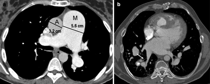
Fig. 18.8
(a) Axial MDTC at the level of the ascending aorta shows a main pulmonary artery (M) to ascending aorta (A) diameter ratio greater than one (PA\AA ratio >1). (b) Axial image shows segmental artery (arrowhead) to bronchus (arrow) ratio >1, better seen at the level of posterobasal segment of the lower lobes
However, the degree of dilation of the main pulmonary artery is not strictly correlated with the mPAP, probably because the caliber of the artery itself depends on many factors, both technical (for instance, the cardiac cycle phase in which the image is obtained), anthropometric (body surface area), or related to diffuse pulmonary disease.
Another finding is a main pulmonary artery-to-aortic diameter ratio greater than one (PA\AA ratio >1), assessed by evaluating the diameter of the main pulmonary artery and of the ascending aorta (Fig. 18.8a). Nevertheless, the elderly may develop a pathophysiological dilation of the ascending aorta, so that this evidence becomes less reliable (Fig. 18.9) (Ng et al. 1999).
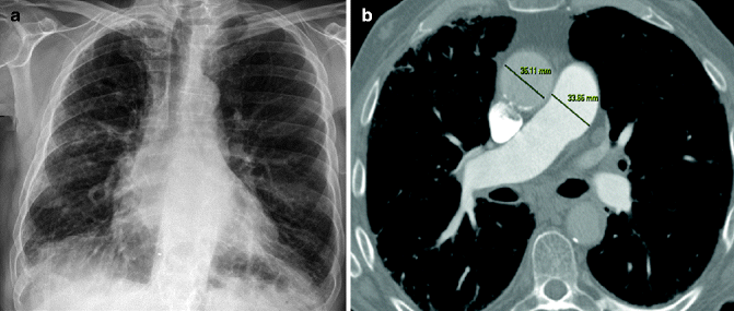
Fig. 18.9
Old patient with PH secondary to COPD. (a) The chest x-ray shows cardiac enlargement, particularly of the right sections and nonspecific signs of COPD. (b) CT demonstrates pulmonary artery enlargement (diameter 33.56 mm). However, the ascending aorta is also dilated (diameter 35.11 mm); consequently the PA\AA ratio is not altered (<1) and unreliable
Hypertrophy of bronchial arteries is another vascular sign of PH visible on CTPA (Fig. 18.10


Related posts:

Stay updated, free articles. Join our Telegram channel
Full access? Get Clinical Tree
