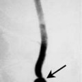
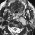
Chapter 125 Amyloid is an extracellular proteinaceous substance consisting of 95% amyloid fibril protein and 5% P component (a glycoprotein). Of the 15 biochemically distinct proteins, the primary components are amyloid light chain (derived from plasma cells) and amyloid-association protein (a unique nonimmunoglobulin synthesized by the liver). Amyloid deposits can be seen in almost all organs and are associated with a diverse number of clinical settings. Amyloidosis may present as a localized lesion or a systemic disease. Systemic amyloidosis may be further classified into primary (associated with immunocyte dyscrasia) and secondary amyloidosis (complication of an underlying chronic disease). Microscopic amyloid deposits in the thyroid gland are not uncommonly seen in systemic disease but localized amyloid goiter is unusual. Thyroid amyloidosis usually presents with a history of painless neck swelling for several months. Clinically, the diffusely enlarged thyroid feels firm, nodular, and nontender. There are usually no associated signs of hyperfunction or hypothyroidism. When amyloid is stained with iodine followed by sulfuric acid it turns blue, a characteristic shared by starch. This observation led Virchow to suggest the term amyloid
Thyroid Amyloidosis
Epidemiology
Clinical Findings
Pathology
![]()
Stay updated, free articles. Join our Telegram channel


Full access? Get Clinical Tree


