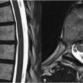
30 Trauma MRI offers superior evaluation of soft tissue and cord injuries and is preferred to CT for this purpose in the setting of trauma. The sagittal fat-saturated T2WI in Fig. 30.1A demonstrates compression of the cervical spinal cord by a small surrounding epidural fluid collection (white arrows). SI characteristics of this lesion on accompanying sequences helped identify it as an epidural hematoma. In the spine, these result from the tearing of the epidural venous plexus. The SI of these hematomas varies with the age of their constituent blood products. A traumatic disk herniation (black arrow) at the C6–C7 level is demonstrated in Figs. 30.1B,C on sagittal FSE T2WI and axial GRE T2WI, respectively. The axial image demonstrates a broad-based central and left paracentral disk herniation (black arrow), with associated mild cord flattening. Traumatic herniations may arise secondary to osseous injury as in the T2WI of Fig. 30.2A. Here the anterior subluxation of C4 relative to C5 has led to a disk protrusion with mild to moderate compression of the cord. A perched facet (white arrow) is also visible on the left parasagittal image in Fig. 30.2B. Occasionally, damage to the cord may occur without direct evidence of concurrent osseous or soft tissue injury. The T2WI of Fig. 30.2C demonstrates longitudinally extending cord edema (black arrows
![]()
Stay updated, free articles. Join our Telegram channel


Full access? Get Clinical Tree


