Traumatic Brain Injury
Ari M. Blitz
Traumatic brain injury (TBI) presenting in isolation or as a component of polyorgan trauma accounts for much of trauma-related morbidity and mortality. Urgent diagnosis and treatment is critical and is most often guided by computed tomography (CT) findings. CT findings which correlate most strongly with poor outcome are subfalcine herniation (also known as midline shift), effacement of basal cisterns (due to edema or herniation), and subarachnoid hemorrhage.
This chapter discusses various forms of traumatic intracranial hemorrhage, as well as other acute injuries such as diffuse axonal injury (DAI), and cerebral contusion. Acute TBI is categorized into extracerebral hemorrhages and intraparenchymal injuries for ease of discussion, although these two forms of injury often coexist. Skull fractures are discussed in Chapter 2, “Skull Fractures”, and patterns of herniation are discussed in Chapter 15, “Herniation”.
Indications.
Because of its speed, availability, and high sensitivity for acute hemorrhage, CT is the primary imaging modality for the assessment of TBI. Magnetic resonance imaging (MRI) is most often used in the subacute stage, and is particularly sensitive to DAI and nonhemorrhagic contusions.
Computed Tomographic Protocol.
Noncontrast head CT reconstructed with both soft tissue and bone algorithms is the imaging standard. Computed tomographic angiography (CTA) may be performed in select patients to look for vascular injuries such as dissection/occlusion and caroticocavernous fistula (CCF).
Magnetic Resonance Imaging Protocol.
Noncontrast MRI evaluation typically includes: sagittal T1, axial T2, fluid attenuated inversion recovery (FLAIR) axial, and diffusion weighted axial with calculated apparent diffusion coefficient (ADC) map. Gradient echo or susceptibility weighted sequences are critical, as they have a high sensitivity for acute and chronic hemorrhage.
Extracerebral hemorrhage may be confined to the epidural space, subdural space, or subarachnoid space. A working knowledge of the anatomy of the meninges and cranial sutures is therefore necessary to appropriately discuss and diagnose intracranial and extra-axial hemorrhage and will often significantly impact upon clinical decision making. Hemorrhage can be superficial to the dura (epidural hemorrhage), subjacent to the dura but superficial to the arachnoid (subdural hemorrhage), and between the arachnoid and the pia mater (subarachnoid hemorrhage).
Epidural Hematoma
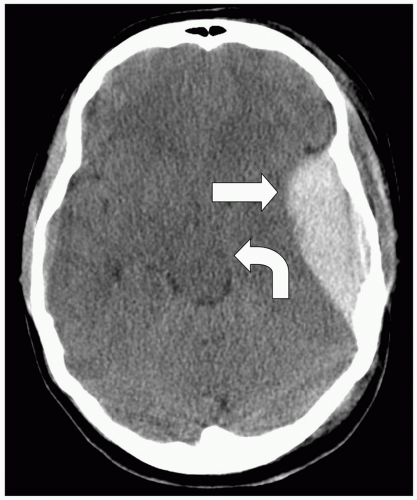
Epidural hematoma (Fig. 1-1) typically occurs in association with a linear fracture of the temporal bone that causes laceration of the middle meningeal artery or one of its branches,
resulting in extravasated arterial blood under high pressure dissecting between the inner table of the skull and the closely applied outer layer of the dura mater. Because epidural hematoma has an arterial source 80% of the time there can be very rapid accumulation of blood, increased intracranial pressure, and subsequent brain herniation. Posterior fossa epidural hematomas are associated with occipital bone fractures and disruption of the transverse sinus. The classic clinical scenario is blunt trauma to the temporal region, loss of consciousness followed by a lucid interval of minutes to a few hours, with subsequent rapid deterioration into coma and even death as the mass effect grows. Epidural hematoma is a neurosurgical emergency.
resulting in extravasated arterial blood under high pressure dissecting between the inner table of the skull and the closely applied outer layer of the dura mater. Because epidural hematoma has an arterial source 80% of the time there can be very rapid accumulation of blood, increased intracranial pressure, and subsequent brain herniation. Posterior fossa epidural hematomas are associated with occipital bone fractures and disruption of the transverse sinus. The classic clinical scenario is blunt trauma to the temporal region, loss of consciousness followed by a lucid interval of minutes to a few hours, with subsequent rapid deterioration into coma and even death as the mass effect grows. Epidural hematoma is a neurosurgical emergency.
 Figure 1-1. Noncontrast head computed tomography (CT) showing lens-shaped left temporal epidural hematoma (straight arrow) causing uncal herniation (curved arrow). |
The dura and bone are essentially fused at sutures, so epidural hematomas do not cross sutures, and therefore take on a characteristic biconvex shape. Epidural hematomas can cross midline.
Stay updated, free articles. Join our Telegram channel

Full access? Get Clinical Tree


