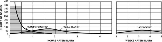
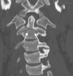
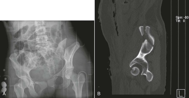
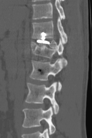
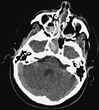
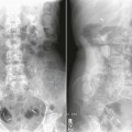
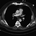
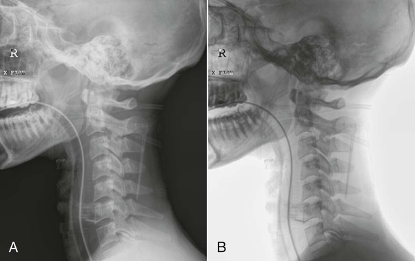
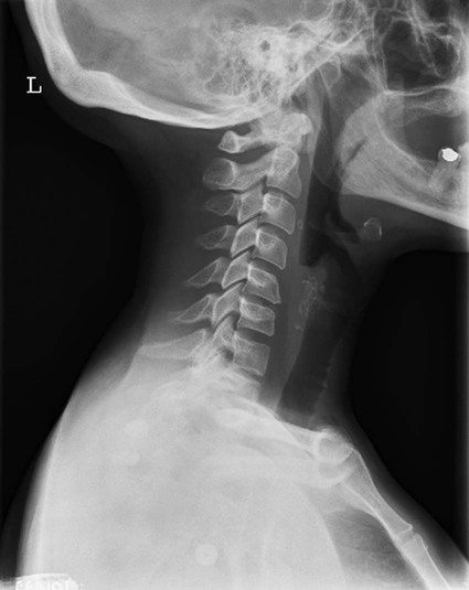
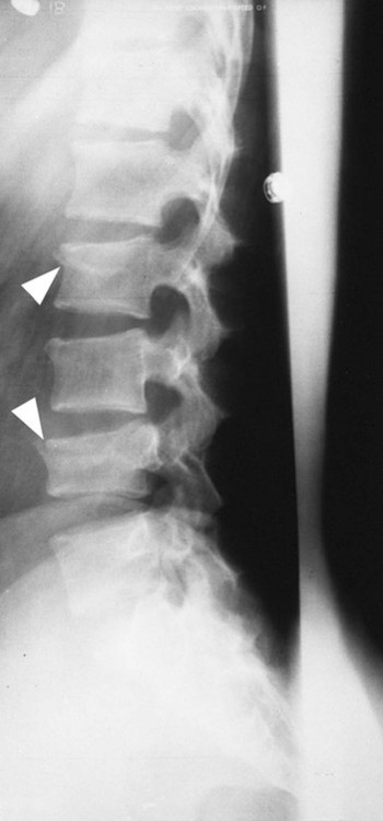
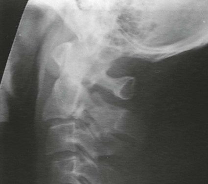
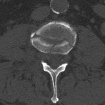
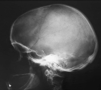
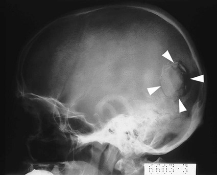
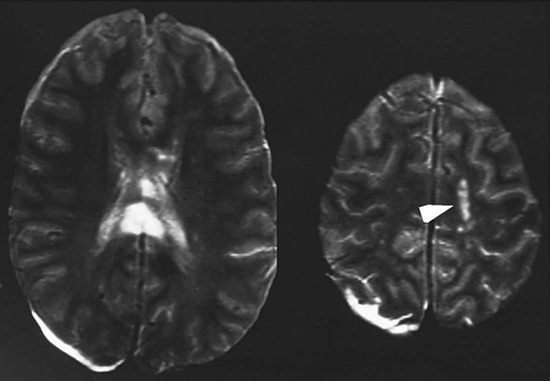
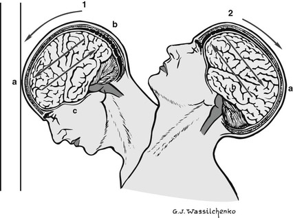
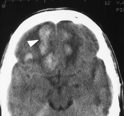
Traumatic Disease


Traumatic Disease


In the United States, injury is one of the leading causes of death and disability, and trauma is the most common cause of death for individuals between the ages of 1 and 44 years, accounting for over 48,000 deaths in 2008. Additional information can be obtained at the Centers for Disease Control and Prevention (CDC) National Center for Health Statistics Web site at www.cdc.gov/nchs/hus.htm. The annual medical cost associated with trauma injury is approximately $200 billion and is classified by several categories. Some examples are as follows:


These costs are a measure of the dollars spent and income not received because of accidents, injuries, or fatalities. These costs also demonstrate the importance of prevention strategies initiated by the National Safety Council.


The American College of Surgeons (ACS) Committee on Trauma periodically reviews its guidelines to ensure optimal patient care by classifying medical centers and hospitals according to their ability to treat various injuries and by publishing guidelines for trauma management. Trauma results primarily from motor vehicle accidents (MVAs), unintentional accidents at home or in the workplace, gunshot wounds, stab wounds, physical altercations, domestic violence, and physical abuse. According to the Centers for Disease Control and Prevention (CDC), hospital emergency departments play a critical role in the U.S. health care system, from treatment of minor trauma to, more recently, the first-line defense against bioterrorism.


Deaths from traumatic injuries have a trimodal distribution (Fig. 12-1), with the first critical period occurring seconds after the injury. Death during this period results from lacerations of the brain and spinal cord or the heart and great vessels. The second critical period occurs during the first 4 hours after the injury, with death generally resulting from intracranial hemorrhage, lacerations of the liver and spleen, or significant blood loss from multiple injuries. The third critical period occurs weeks after the injury, when death results from infection and multiple organ failure.


A well-designed emergency medical system (EMS) provides for prehospital care, acute hospital care, and rehabilitative care. Medical facilities are classified as level I, level II, or level III trauma centers based on the availability of specialized medical personnel and equipment. When patients are triaged (medically screened to determine their relative priority for treatment), at the site of an accident, their injuries are classified as life-threatening, urgent, or nonemergent. Multiple injuries most likely occur in conjunction with severe head injuries. These patients must be treated with utmost care. Before a trauma victim is transported, a clear airway must be established, acute bleeding must be controlled, and the patient must be immobilized to avoid displacing fractures of the skeletal system. Immobilization of the spine is paramount to avoiding further injury to the spinal cord, and this is accomplished with the use of splints, backboards with head blocks, cervical collars, and special air splint suits.


The EMS ensures that the trauma victim is taken to the closest appropriate medical facility to receive the proper medical care for the injuries, but not necessarily to the closest hospital. Once the patient arrives at the proper medical facility, another careful assessment is necessary. This secondary triage assessment includes evaluation of the patient’s state of consciousness, vital signs (blood pressure, pulse, temperature, and respiration), pupil size and reaction to light, and motor activity of the extremities. Cross-table lateral cervical spine radiography or computed tomography (CT) of the cervical spine is necessary to assess any damage to the cervical spine before the patient is moved. In addition, radiographs of the chest, abdomen, and skeletal system must be obtained to evaluate the extent of injuries. Although national vital statistics show that only 5% of all trauma victims have life-threatening injuries, these types of injuries are responsible for 50% of all in-hospital trauma-related deaths.


Level I, II, and III Trauma Centers


The primary hospitals in the trauma system are level I trauma centers. Such medical centers can provide total care for all injuries. Key elements of a level I trauma center include 24-hour in-house coverage by general surgeons and prompt availability of care in specialties such as orthopedic surgery, neurosurgery, anesthesiology, emergency medicine, radiology, internal medicine, and critical care. Other capabilities include cardiac, hand, pediatric, and microvascular surgeries and hemodialysis. The level I trauma center provides leadership in prevention, public education, and continuing education of the trauma team members, as well as serving as a research facility to help direct new innovations in trauma care. Level I trauma centers are generally located in large metropolitan areas and serve as both primary care and tertiary care institutions. Another stipulation for a health care institution to be classified as a level I trauma center is that it must treat 1200 admissions or 240 major trauma patients per year.


Level II trauma centers are the most common trauma facilities serving as community trauma centers. These institutions can handle the majority of trauma cases and transport patients to level I facilities only when necessary. Level II facilities include 24-hour immediate coverage by general surgeons, as well as coverage by the specialties of orthopedic surgery, neurosurgery, anesthesiology, emergency medicine, radiology, and critical care. More critical patients with needs such as cardiac surgery, hemodialysis, or microvascular surgery may be referred to a level I trauma center. These medical centers are generally community hospitals located in smaller cities and towns and provide a valuable service.


Level III trauma centers are usually located in remote rural areas and serve communities that do not have a level II center. A level III trauma center has a demonstrated ability to provide prompt assessment, resuscitation, stabilization of injured patients, and emergency operations. The key components of a level III trauma center include 24-hour immediate coverage by emergency medicine physicians and the prompt availability of general surgeons and anesthesiologists. The level III trauma center has formal transfer agreements for patients requiring more comprehensive care at a level I or level II trauma center.


As determined by the ACS Committee on Trauma, a level IV trauma center has a demonstrated ability to provide advanced trauma life support (ATLS) prior to transfer of patients to a higher-level trauma center. A level IV trauma center has basic emergency department facilities to implement ATLS protocols and 24-hour laboratory coverage. Transfer to higher-level trauma centers follows the guidelines outlined in formal transfer agreements.


Imaging Considerations


Radiography


Conventional radiography is one of the first imaging modalities required in the management of a trauma victim. A cross-table lateral cervical spine radiograph may be obtained to evaluate the presence or absence of a fracture before the patient is moved. Portable chest, abdominal, and pelvic radiographs are also generally obtained as soon as the patient arrives in the emergency department.


Conventional radiography is still the primary means of evaluating skeletal trauma. Additional information about damage to the muscle, tendons, ligaments, and soft tissue structures is obtained using magnetic resonance imaging (MRI). CT and nuclear medicine studies may also be employed to identify subtle skeletal fractures.


Computed Tomography


The high-energy impact associated with most MVAs often results in injury to the neck and head, so CT is an important tool in assessing the trauma victim. CT is excellent for imaging acute cerebral hemorrhage and fractures of the skull and facial bones. Guidelines governing the use of CT in the case of head trauma include the Canadian rule, which suggests that injuries with a Glasgow Coma Scale (GCS) score of 13 to 15 should be evaluated by CT, as well as the more comprehensive New Orleans helical computed tomography (HCT) rule. The New Orleans HCT rule mandates CT examination of patients with a GCS score of 15 in combination with any one of the following: headache, vomiting, age above 60 years, drug or alcohol intoxication, persistent amnesia, visible trauma above the clavicle, or seizure. In lieu of the increased awareness of radiation dose, patient protection, and public safety campaigns such as Image Gently and Image Wisely, the ACR Appropriateness Criteria now include a relative radiation level (RRL) for imaging procedures for both adult and pediatric patients.


With the prevalent use of multidetector CT (MDCT), CT evaluation of the cervical spine has become a standard procedure in evaluating trauma patients. The National Emergency X-radiography Utilization Study (NEXUS) and Canadian C-Spine Review (CCR) concluded that CT demonstrates 99.6% sensitivity for the identification of cervical spine fractures. CT has replaced conventional radiography as the first-line approach to imaging the cervical spine, with the meta-analysis of research studies performed in 2005 indicating that CT demonstrates a much higher specificity of 98% compared with a specificity of 52% for conventional radiography of the cervical spine. Axial CT of the cervical spine is performed in conjunction with coronal and sagittal reconstruction to fully assess the areas of interest (Fig. 12-2). With more sensitive imaging techniques now available, CT and MRI have revealed a significant number of fractures and other injuries that are not diagnosed with radiography. CT evaluation can reduce hospital admission rates and results in more efficient surgical intervention by accurately identifying the presence and extent of injury in the trauma patient.


Blunt trauma to the abdomen is best evaluated with CT or abdominal sonography. Trauma to the urinary system occurs in about 10% to 15% of patients who experience blunt trauma to the lower abdomen and pelvis. In cases of hematuria, CT is preferred to conventional intravenous pyelography (IVP) or intravenous urography (IVU). Although the use of oral contrast agents for abdominal CT is debatable, intravenous contrast is routinely employed to allow evaluation of vascular injuries and to better visualize the spleen, pancreas, and kidneys. In addition, in cases of blunt abdominal trauma, CT is better able to visualize fractures of the transverse processes of the lumbar spine, often missed on conventional spine radiography. CT, in combination with a conventional anteroposterior (AP) pelvic radiography, is also used to assess pelvic fractures commonly associated with abdominal injury (Fig. 12-3, A). In cases of pelvic fractures, it is also imperative to identify any possible trauma to the urinary bladder and to perform CT (see Fig. 12-3, B) to determine the extent of injury. Occasionally, emergent cystography may be performed to visualize any injury to the urinary bladder. In cases of penetrating trauma to the abdomen, angiography may also be used to identify the extent of injury. Sonography plays little or no role in the evaluation of genitourinary trauma. Several studies have documented the inability of sonography to detect injuries of the kidney or bladder in trauma patients.


Trauma of the Vertebral Column and Head


The initial management of patients with head and spinal trauma is critical. Key items to assess at the scene of the accident include the mechanism of injury and changes in the patient’s neurologic status. A neurologic assessment using the GCS should be conducted at the scene, and cervical spine injury should be assumed to be present until it is ruled out by radiologic investigation. A cervical spine fracture can be present in up to 20% of patients with a severe head injury.


The head and neck should be immobilized, and care must be taken if intubation is necessary. Hyperextension injuries of the head and neck or direct trauma to the neck may injure the carotid arteries. Bleeding must be controlled to prevent shock, which may worsen the head injury. Once the patient arrives in the emergency department, cervical spine radiographs or a CT scan of the cervical spine must be obtained. CT of the head may also be indicated, especially if the patient is comatose.


Many studies have been conducted regarding the effectiveness of airbags and seatbelts in preventing head and neck fractures in the event of an MVA. By far, those who do not use any protective device sustain the most injuries. Those who depend only on the airbags sustain the second largest number of injuries to the head and face, followed by those who use only a seatbelt. The most effective protection results from the use of both protective devices. In addition, individuals using only a lap-type seatbelt have a high incidence of lumbar spine injuries, and individuals wearing only a shoulder belt without a lap belt sustain more cervical spine injuries.


Injuries to the Vertebral Column


The causes of vertebral column injuries include direct trauma and hyperextension–flexion injuries (whiplash injuries). Radiographic indications of spinal column injuries include the interruption of smooth, continuous lines formed by the vertebrae stacked on one another (Fig. 12-4). Either a dull or sharp pain in the posterior neck is the primary manifestation of a whiplash injury. The pain may radiate down the arms or back. Muscle spasm as a result of trauma may cause a reversal or straightening of the normal spine curvatures. Imaging of whiplash injuries is limited to soft tissue studies, with the exclusion of fractures and dislocations being the first priority. The loss of lordosis is the most common finding on radiographs of patients with whiplash injuries (Fig. 12-5).


Perhaps the most common condition of the vertebral column is generalized back pain, typically in the lumbar area. This may result from injury to the area or from degenerative disease. Such back pain may not always result from bone involvement. Disk disease may cause muscle spasm, with pain referral throughout the back or down the legs. Finally, back pain may be secondary to referred pain from the hip.


Compression fractures are the most frequent type of injury involving a vertebral body. Usually, the damage is limited to the upper portion of the vertebral body, particularly to the anterior margin. Such fractures generally occur in the thoracic and lumbar vertebrae (Fig. 12-6), with the most common site being T11-T12 in the thoracic spine and T12-L1 at the thoracolumbar juncture. Compression fractures are also associated with osteoporosis and range from mild to severe. More severe fractures may cause significant pain, leading to inability to perform activities of daily living to life-threatening decline in the older patient. Cervical spine injuries may involve the odontoid process, usually at the junction of the odontoid and the body of the second cervical vertebra (see Fig. 12-2). A hangman’s fracture (Fig. 12-7) is a fracture of the arch of the second cervical vertebra and is usually accompanied by anterior subluxation of the second cervical vertebra on the third cervical vertebra. A hangman’s fracture, sometimes referred to as traumatic spondylosis, results from acute hyperextension of the head.


Jefferson fracture was first described as a “burst fracture” of the first cervical vertebra (atlas). It generally occurs as a result of a severe axial force that causes compression, as in a diving accident. The vertebral arch literally bursts. Radiographically, particular attention needs to be paid to the transverse longitudinal ligament by reviewing the lateral masses on the open-mouth odontoid projection. MRI is the preferred imaging modality to best examine the transverse longitudinal ligament.


Radiography or CT of the trauma patient with vertebral trauma is critical. Statistically, 5% to 10% of patients with one spinal fracture have another fracture elsewhere in the vertebral column. Fractures and dislocations of the spine are classified as stable or unstable. The spine may be visualized as two columns, with the anterior column composed of vertebral bodies and intervertebral disks and the posterior column composed of the posterior elements (e.g., spinous processes, lamina). If either the anterior column or the posterior column of the spine is fractured or dislocated, the injury is classified as stable. However, if both columns are involved in the injury, it is classified as unstable. In all cases, the patient should be immobilized until cross-table lateral radiographs or CT images have been obtained and cleared by a physician. To rule out possible fractures and dislocations, lateral cervical spine radiography must include all seven vertebrae in their entirety, including spinous processes and intervertebral disk spaces. At times, this may require assistance in depressing the patient’s shoulders or the use of the specialized cervicothoracic lateral projection (Twining or swimmer’s method) to clearly demonstrate the entire seventh cervical vertebra and the upper thoracic vertebra in a lateral projection. Additional trauma projections of the cervical spine such as the pillar projection or trauma oblique projection may be requested to better demonstrate the complex anatomy of the spine. Trauma cervical spine radiographs are analyzed to evaluate (1) the size, shape, and alignment of the vertebral bodies and spinous processes, (2) the position and integrity of the odontoid process of C2, (3) the orientation and clarity of the facet joints, (4) the relationship of C1 to the occipital bone, (5) the alignment of the spinolaminal lines, and (6) any prevertebral swelling.


Spinal injury often results in a loss of neurologic function that may be temporary or permanent, depending on the cause of the dysfunction. Compression of the spinal cord by contusion or hemorrhage leads to rapid swelling of the spinal cord. This causes a rise in intradural pressure and causes temporary neurologic dysfunction. This temporary loss of neurologic function usually resolves in several days. However, lacerations of the spinal cord or transection of the cord results in permanent damage because the severed nerves do not regenerate. Laceration of the spinal cord above the fifth cervical vertebra is almost always fatal, and lacerations below this region result in permanent paralysis. Patients with lacerations or transection of the cord develop immediate flaccid paralysis with loss of all sensation and reflex activity, which gradually changes to spastic paraplegia within days.


Fractures or dislocations of the vertebrae may impinge on the spinal cord and cause significant damage. The responsibility of the radiographer in terms of proper patient handling and obtaining diagnostic-quality images cannot be overemphasized. CT plays a vital role in the diagnosis and treatment of vertebral fractures, dislocations, and associated problems (Fig. 12-8). In certain situations, MRI may be used to evaluate the extent of ligamentous and soft tissue injury or injury to the spinal cord.


Stable injuries to the spine are treated with complete bed rest and steroids until swelling and pain subside. Unstable injuries are immobilized with traction until bone and soft tissue structures have healed. Cervical radiographs are often obtained to demonstrate proper alignment while the patient is in traction. Surgery may also be necessary for internal fixation of the fractures or to remove displaced fragments and decompress the spinal cord. CT of the spine is used preoperatively to serve as a road map for the surgeon because this imaging modality clearly demonstrates the size, number, and location of various fracture fragments. It may also be used postoperatively to demonstrate the success of the surgical procedure (Fig. 12-9).


Injuries to the Skull and Brain


The anatomy surrounding the delicate brain generally protects it well under normal conditions. The diploic arrangement of the calvaria, the mechanical buffering action of cerebrospinal fluid, and the tough dura mater all work to prevent brain injury. Despite this protection, sufficient force to the skull may cause injury to the brain. Head trauma is the major neurologic cause of mortality and morbidity in individuals under 50 years of age.


Head trauma may result in skull fractures, brain injury, or a combination of the two. The role of plain film radiography in evaluation of head trauma is rather limited, but CT allows rapid assessment of the nature of any brain injury. Assessment of the state of the brain after head injury is more crucial than that of the skull. For trauma victims, routine skull radiography may be delayed to allow treatment of the complications of brain injury readily diagnosed by CT. Skull fractures visualized by either modality are often seen with accompanying hematomas. If patients sustain an open skull fracture, they are at risk for development of meningitis or brain abscesses. Regardless of the imaging modality used, the technologist must constantly observe a patient with a head injury while performing an examination. Any change noted in the patient’s condition should be reported immediately.


Cerebral Cranial Fractures.


The term cerebral cranial fractures usually refers to fractures in the calvaria of the skull. Vascular markings in the skull, either venous or arterial, are routinely demonstrated as linear translucencies and may occasionally be mistaken for cerebral cranial fractures (Fig. 12-10). In most cases, a fracture appears more translucent than a vascular marking because a fracture traverses the full thickness of the skull. Although the edges of the fractures may branch abruptly, they may be seen to fit together, whereas venous channels have irregular edges that cannot be fitted together. The sutures between the individual cranial bones remain visible radiographically, even after they become fused. To an untrained eye these sutures may also resemble a fracture.


In most cases, the location of the skull fracture is more important than the extent of the fracture. If the fracture crosses an artery, an arterial bleed may occur, resulting in an epidural hematoma. A fracture that enters the mastoid air cells or a sinus communicates with a potentially infected space and may lead to infection, possibly resulting in encephalitis or meningitis.


Fractures visible after skull trauma are generally classified as linear, depressed, or basilar skull fractures. Linear fractures appear as straight, sharply defined, nonbranching lines and are intensely radiolucent (Fig. 12-11). Up to 80% of all skull fractures are linear fractures. A depressed fracture appears as a curvilinear density because the fracture edges overlap (Fig. 12-12). These fractures are caused by high-velocity impact by small objects. Injury to the cerebral cortex may result, causing bleeding into the subarachnoid space. A depressed fracture is best demonstrated when the x-ray beam is directed tangentially to the fracture.


Basilar skull fractures are very difficult to demonstrate radiographically. The presence of air–fluid levels in the sphenoid sinus or clouding of the mastoid air cells is often the only radiographic finding suggesting a fracture. Therefore, it is important to include cross-table lateral skull radiography with the trauma skull radiographic series. CT and MRI are often used to better identify basilar area fractures and associated soft tissue damage within the skull (Fig. 12-13).


Brain Trauma.


In addition to brain injury from a penetration wound (as could happen with a fracture), brain injury may also occur from acceleration and rapid deceleration of the head, which is termed a closed head injury or traumatic brain injury (TBI). With head trauma, the brain is traumatically shaken within the cranium and subjected to forces of compression, acceleration, and deceleration. Brain tissues are injured from compression, tension, and shearing, with the last perhaps most important (Fig. 12-14). The superficial cerebrum in the frontal, temporal, and occipital regions is most often affected.


TBI is a serious public health problem in the United States. Each year, traumatic brain injuries contribute to a substantial number of deaths and cases of permanent disability. Recent data from the CDC show that, on average, approximately 1.7 million people sustain a TBI annually. This incidence has increased significantly with athletics-related activities, and ongoing research is focusing on athletes from the junior high to professional levels.


After a blow to the head, an individual may experience temporary loss of consciousness and reflexes. This widespread paralysis of brain function is known as a concussion and is characterized by headache, vertigo, and vomiting. Higher mental functions may be impaired for several hours, with the patient remembering little of the events surrounding the concussion. A strong tendency toward spontaneous and complete recovery exists because no structural damage to the brain occurs. Recovery generally takes place in less than 24 hours. Treatment is conservative once assessment (usually by CT) has ruled out any hemorrhage or fracture. Bed rest and possible admission to the hospital are the usual means of dealing with concussion.


A brain contusion may also result from a direct blow to the head. This bruising of brain parenchyma is more serious than a concussion. A contusion formed on the side of the head where the trauma occurs is called a coup lesion, and one formed on the opposite side of the skull in reference to the site of trauma is a contrecoup lesion (Fig. 12-15). Contusions are characterized by neuron damage, edema, and punctate (pinpoint punctures or depressions) hemorrhaging. On CT, contusions appear as small, ill-defined foci of increased density (Fig. 12-16). Subdural or epidural hematomas may occur in conjunction with a contusion and result in increased intracranial pressure that may be life-threatening. Signs seen in the patient with a contusion include drowsiness, confusion, and agitation. Hemiparesis and unequal pupil size may also be seen. CT plays a major role in the diagnosis of hematomas resulting from contusions, providing ready visualization of hemorrhagic blood, as described in the following section. Treatment is generally conservative, centering on prevention of shock, control of edema, and drainage of any hematoma present.


Persistence of loss of consciousness for more than 24 hours is known as a coma. This is usually a serious condition and may be fatal. Comas result from trauma to the head as well as from nontraumatic metabolic malfunctions or circulatory problems that prevent sufficient blood flow to the brain. Diagnosis related to a coma may involve use of CT, MRI, positron emission tomography (PET), or fusion imaging and focuses on determining, if possible, the cause of the coma. Treatment then rests on success in treating and alleviating the cause of the coma.


Hematomas of the Brain.


As noted earlier, brain trauma may result in hemorrhaging of blood from a ruptured artery or vein. Although venous bleeding occurs more slowly than arterial bleeding, both types of hemorrhaging and resultant edema of the brain cause an increase in intracranial pressure. Because the skull’s structure does not allow expansion, the increased pressure displaces the brain toward its opening, the foramen magnum. This trauma to the brain results in serious neurologic consequences or even death if not treated promptly. CT plays the major imaging role in diagnosis of the hemorrhaging.


A hematoma is a collection of blood; four primary types of cerebral hematomas have been identified: epidural, subdural (Fig. 12-17), subarachnoid, and intracerebral. The highest mortality rate is associated with an epidural (extradural) hematoma (Fig. 12-18). Even when promptly recognized and treated, it has a mortality rate of up to 30%. An epidural hematoma results from a torn artery, usually the middle meningeal artery, with blood pooling between the bony skull and the dura mater. Most commonly, the artery or its branches are torn by a fracture of the thin, squamous portion of temporal bone. In more than 80% of cases, the skull fracture is visible radiographically. As an arterial bleed, it accumulates rapidly and quickly causes neurologic symptoms, including early coma. It is seen on CT scans as an increased density, generally occupying a small area with a sharply convex appearance. It is often accompanied by a fracture of the skull or facial bones. If not diagnosed and surgically treated quickly, the outcome is fatal as a result of brain displacement and herniation.

Only gold members can continue reading. Log In or Register to continue

Stay updated, free articles. Join our Telegram channel

Mar 6, 2016 | Posted by in GENERAL RADIOLOGY | Comments Off on Traumatic Disease

Full access? Get Clinical Tree

Get Clinical Tree app for offline access