The interventional suite plays host to a wide variety of patient types and procedures. Some of the patients who arrive in the suite are in good baseline health, others are severely ill. In either case, patients can experience a life-threatening emergency during any interventional procedure. Unlike the operating room, where an anesthesiologist is routinely present to handle any non–procedure-related emergency, the level of support in the interventional suite is variable. When medical emergencies occur, it is not uncommon for the interventionalist to have only their complement of nurses and technicians as support staff. In this brief chapter, we endeavor to provide a framework that will help mitigate the medical emergencies that can occur in patients undergoing interventional procedures.
The lion’s share of medical emergencies can be placed into five categories: (1) oversedation, (2) airway compromise, (3) respiratory distress, (4) cardiac/hemodynamic emergencies, and (5) contrast reactions. The response to these medical emergencies often requires an anesthesiologist, an intensivist, and/or a surgeon. However, there are concrete steps and planning that can help mitigate these emergencies by the interventionalist and their staff, which can prove lifesaving when appropriate staff and resources are mobilized. This chapter focuses on these bridging and mitigating techniques.
Oversedation
Procedural sedation and analgesia is a common and important component of interventional procedures. The appropriate positioning of the patient and maintaining patient comfort is facilitated by proper sedation. However, reaching the perfect balance of sedation and comfort without oversedating the patient can be challenging. Striking the right balance of sedation can be particularly difficult in patients with underlying lung disease and in patients with unpredictable metabolism of common sedatives. In addition, patients with a marginal circulatory status may be prone to hemodynamic effects. The oversedated patient can experience hypercarbia, hypoxemia, and/or hypotension related to the sedative itself, or due to secondary effects. Oversedation and hypercarbia can be prevented with close monitoring of the patient and the use of pulse oximetry; patient’s response to verbal commands; observation of ventilation, blood pressure, and heart rate every 5 minutes; electrocardiography for patients with significant cardiovascular disease; and the use of exhaled capnography. When patients become oversedated, it is common for the patient’s minute ventilation to decrease, which is often associated with a rise in the end-tidal CO 2 . This elevation typically precedes apnea and can alert the physician that the level of sedation is excessive. This is vitally important because many patients have a variable response to hypnotic drugs.
End-tidal CO 2 >50 mm Hg, an absolute change of >10 mm Hg, or an absent waveform may detect subclinical respiratory depression not detected by pulse oximetry alone. And capnography is 18 times more likely to detect respiratory depression compared with pulse oximetry.
The choice of drugs that have minimal hemodynamic and respiratory depression may be considered; such drugs include etomidate, ketamine, ketamine-propofol combination, and dexmedetomidine.
In this section, we focus on the airway and respiratory management of the oversedated patient. The management of hypotension and arrhythmia is covered in the circulatory management section of this chapter.
When patients become oversedated, their airway and respiratory centers can be compromised. The mechanism by which this occurs are multiple: (1) use of narcotics can cause vomiting and aspiration, (2) narcotic and benzodiazepine use decreases respiratory drive and can cause central apnea, (3) muscle relaxation caused by sedatives can cause upper airway obstruction. Typically, the response to the oversedated patient is to stop administration of the sedative agent, increase inspired oxygen (e.g., placing a 100% fraction of inspired oxygen face mask), and start some form of stimulation (e.g., sternal rub) in an attempt to arouse the patient to stimulate breathing while the sedative wears off. This approach is often not satisfactory because of the half-life of the sedating agent. In addition, if the airway or respiratory center is compromised, the subsequent hypercarbia may spiral toward complete respiratory arrest.
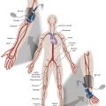
The approach to the oversedated patient should be managed in a more airway-centered manner. Once oversedation is recognized, a staff member in the interventional suite should be detailed to the head of the bed to manage the airway. Noninvasive airway management should be sufficient in most cases. If stimulation of the patient (e.g., calling the patient’s name loudly, performing sternal rub) fails to immediately correct the compromise in oxygenation or respiratory effort, the airway should be assessed and optimized (see the Airway Management and Optimization section and Fig. 4.1 ). At this point, if the sedative used has an antidote (e.g., flumazenil for benzodiazepines or naloxone for narcotics), that antidote should be drawn up ( Table 4.1 ).

| Antidote | Dosage and Comments | Side Effects |
|---|---|---|
| Naloxone | 0.4–2 mg at a time; maximum dose 10 mg For typical doses used for conscious sedation, a dose of 0.2–0.4 mg should be sufficient. Can be given as an infusion (2 mg/100 mL) 0.1 mg/min. | As narcotic reverses, elevated blood pressure, tachycardia, nausea, and vomiting can occur. |
| Flumazenil | 0.2–1 mg dose Each 0.2-mg increment should be given over 1 min. In clinical trials, 75% of patients responded to a dose of 1–3 mg. | Frequent (≈10%): nausea, vomiting, headache, blurred vision Major (<1%): seizure |
Optimization of the airway often requires repositioning the patient’s head and neck. A simple understanding of the anatomy allows anyone to place the patient’s head in more favorable position for gas exchange. The tongue is the most common cause of upper airway obstruction. A head-tilt/chin-lift or jaw-thrust maneuver ( Fig. 4.2 ), also called the sniffing position, may establish an airway. If, however, proper positioning fails to establish a viable airway, more invasive maneuvers such as using a nasopharyngeal/oropharyngeal airway or a laryngeal mask airway may be needed. These are be discussed in a later section.

Both benzodiazepines and narcotics have reversal agents that if used correctly could save an oversedated patient from having a respiratory arrest. Flumazenil antagonizes the action of benzodiazepines on the central nervous system and inhibits activity at γ-aminobutyric acid/benzodiazepine receptor sites. It is contraindicated in patients with serious tricyclic overdoses. Available in injectable 0.1 mg/mL, flumazenil can be given undiluted or in 5% dextrose in water, lactated Ringer’s solution, or or 0.9% normal saline. The initial dose is 0.2–0.4 mg intravenously (IV) over 15 seconds. If ineffective, a second dose can be given every 60 seconds. Maximum initial dose is 1.0 mg. If the patient becomes oversedated, an additional dose can be given and a drip may need to be started because the half-life of the offending benzodiazepine may be longer than the half-life of flumazenil, whose half-life is 40–80 minutes. Adverse effects include seizures, dizziness, headache, blurred vision, diplopia, visual field deficit, hyperventilation, nausea, and vomiting.
Naloxone hydrochloride is a pure narcotic antagonist. It reverses the respiratory depression, sedation, and hypotensive effects of opioids. In the absence of narcotics, naloxone has no activity. Caution needs to be used in narcotic addiction, cardiac disease, and use of cardiotoxic drugs. The rapid reversal of narcotic depression may also cause nausea, vomiting, diaphoresis, and circulatory stress. The initial dose is 0.1–2mg IV at 2–3-minute intervals until reaching desired effect of reversal. It can be given undiluted as a bolus or as an IV infusion with 5% dextrose in water or 0.9% normal saline. As in flumazenil a drip may need to be started due to a longer half-life of the offending agent. Naloxone has a half-life of 30–81 minutes. As an infusion, use 3.7 μg/kg/h. Adverse effects include seizures, ventricular tachycardia, ventricular fibrillation, acute narcotic abstinence syndrome, and return of pain.
If stimulation, airway optimization, and reversal agents fail to rectify the situation, airway compromise may be the underlying problem and then the clinician should continue down the airway management algorithm (see Fig. 4.1 ) .
Airway Management and Optimization
Airway management is a core skill for the medical specialties of anesthesiology, critical care medicine, and emergency medicine. A small section in a chapter is not sufficient to cover all aspects of this topic. In this section, the goal is to arm the interventionalist with some basic noninvasive skills and maneuvers that can mitigate an impending airway calamity. As mentioned earlier, we recommend that some members of the interventional support staff have some basic noninvasive airway management training. Advanced cardiac lifesaving (ACLS) training for all interventional nurses would be optimal, but this may not be a reasonable request given overall staffing considerations.
There are a variety of noninvasive airway management maneuvers that can dramatically improve ventilation and oxygenation pending a more durable intervention (e.g., endotracheal intubation). Suctioning, head positioning, and keeping a patent airway are the first and often highest-yield steps.
- 1.
The oropharynx should be suctioned under direct vision in order to remove any secretion accumulation. This can be diagnostic and therapeutic.
- 2.
The head position should be adjusted in order to maximize airway patency (see Fig. 4.2 ). The sniffing position mentioned previously with the head-tilt/chin-lift and/or jaw-thrust maneuver should be performed to optimize the airway (see Fig. 4.2 ).
- 3.
If these maneuvers do not rectify the situation:
- •
A nasopharyngeal airway should be placed. This is a noxious stimulus and the act of placing it may reestablish the airway. The nasopharyngeal airway must have lubricant applied at its end. It is placed in either naris until it is hubbed. Once applied, bag-valve masking should be reinstituted. The adequacy of bag-valve masking can be assessed by pulse oximetry and also by the rise of the thorax with each breath delivered by the bag-valve mask.
- •
If bag-valve masking with a nasopharyngeal airway in place fails to correct the hypoxia and/or hypoventilation, a laryngeal mask airway should then be considered and a rapid response team or a code should be called.
- •
- 1.
Airway Compromise
After the above steps have failed, the next immediate step is using a bag-valve mask to breathe for the patient with the understanding that a respiratory arrest may be imminent. Following a simple algorithm may prevent oversedation from becoming airway compromise and ultimately respiratory arrest. The interventionalist should feel comfortable with a certain sequence of events when a patient becomes unresponsive ( Fig. 4.1 ). Initial management should always be stimulation, airway optimization, use of reversal agents where appropriate, and, finally, use of a bag-valve mask. If bag-valve ventilation does not result in immediate resolution of hypoxemia, a code should be called, and ACLS protocols should be initiated. Fortunately, this extreme level of intervention is not commonly required. The simple skill set of breathing for a patient can prevent airway compromise from becoming a code. It is important to have a member of the interventional team who has ACLS certification. If full ACLS certification is not possible, it takes an afternoon of time with an anesthesiologist to learn how to effectively use a bag-valve mask to breathe for a patient. Taking the time to train some of the interventional support staff to have some proficiency around the airway is an excellent investment in preventative medicine even for those staff members who are already ACLS-certified. The interventional suite should have the following materials at all times:
- •
suction set-up
- •
access to high-flow oxygen
- •
oral and nasopharyngeal airways
- •
pulse oximetry
- •
ability to assess blood pressure and respiratory effort
- •
the reversal drugs naloxone and flumazenil
- •
bag-valve mask (also called “ambu bags”)
- •
laryngeal mask airways in various sizes
- •
a stethoscope
- •
In our experience of managing procedural sedation for a variety of different procedures, mask breathing for the patient while the sedative wears off to a more appropriate level is adequate therapy for the oversedated patient in most cases. Table 4.2 outlines the contingency kit that should be immediately available for all interventional procedures.
Related posts:

Stay updated, free articles. Join our Telegram channel

Full access? Get Clinical Tree


