, Joungho Han2, Man Pyo Chung3 and Yeon Joo Jeong4
(1)
Department of Radiology Samsung Medical Center, Sungkyunkwan University School of Medicine, Seoul, Korea, Republic of (South Korea)
(2)
Department of Pathology Samsung Medical Center, Sungkyunkwan University School of Medicine, Seoul, Korea, Republic of (South Korea)
(3)
Department of Medicine Division of Pulmonary and Critical Care Samsung Medical Center, Sungkyunkwan University School of Medicine, Seoul, Korea, Republic of (South Korea)
(4)
Department of Radiology, Pusan National University Hospital, Busan, Korea, Republic of (South Korea)
Abstract
Tree-in-bud sign refers to the condition in which small centrilobular nodules less than 10 mm in diameter are associated with centrilobular branching nodular structures [1] (Fig. 9.1). The small nodules represent lesions involving the small airways. However, vascular lesions involving the arterioles and capillaries may simulate the centrilobular small nodules and branching nodular structures (vascular tree-in-bud sign) [2–5] (Fig. 9.2).
Definition
Tree-in-bud sign refers to the condition in which small centrilobular nodules less than 10 mm in diameter are associated with centrilobular branching nodular structures [1] (Fig. 9.1). The small nodules represent lesions involving the small airways. However, vascular lesions involving the arterioles and capillaries may simulate the centrilobular small nodules and branching nodular structures (vascular tree-in-bud sign) [2–5] (Fig. 9.2).
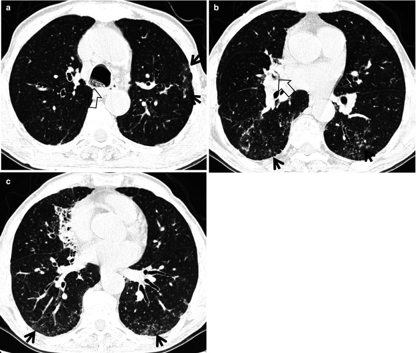
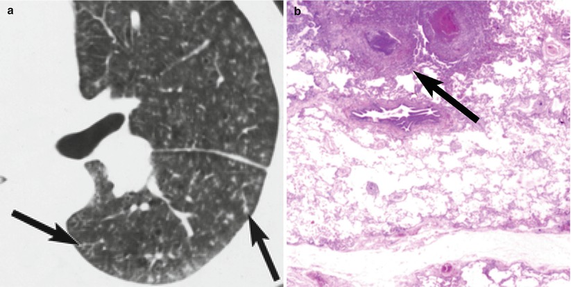
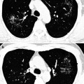
Fig. 9.1
Aspiration pneumonia in a 66-year-old man with Parkinson’s disease. (a–c) Lung window images of thin-section (1.0-mm section thickness) CT scans obtained at levels of the azygos arch (a), right middle lobar bronchus (b), and inferior pulmonary veins (c), respectively, show multifocal areas of tree-in-bud signs (arrows) in both lungs and parenchymal consolidation in the right middle lobe. Also note large amount of secretion (open arrows) in tracheobronchial trees.
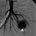
Fig. 9.2
Cellulose granulomatosis (foreign-body-induced pulmonary vasculitis) in a 37-year-old man who is a drug abuser. (a) Thin-section (1.5-mm collimation) CT scan obtained at level of left interlobar pulmonary artery shows diffuse pulmonary involvement with ill-defined centrilobular small nodules (arrows). Note also nodular branching structures (tree-in-bud sign). (b) Low-magnification (×40) photomicrograph demonstrates necrotizing vasculitis at the central portion of secondary pulmonary lobule (arrow) (Reprinted from Han et al. [3] with permission)
Diseases Causing the Sign
The localized tree-in-bud sign is typically seen in infectious bronchiolitis, aspiration bronchiolitis, and bronchopneumonia including tuberculosis and nontuberculous mycobacterial pulmonary disease. Vascular tree-in-bud sign can be observed in foreign-body-induced necrotizing pulmonary vasculitis (cellulose and talc granulomatosis) [2–4] and in localized pulmonary lymphatic metastasis [5] (Table 9.1). For diseases related to this chapter, please refer to section “Small Nodules with Centrilobular Distribution” in Chap. 18 and Chap. 26.
Table 9.1
Common diseases manifesting as tree-in-bud sign
Disease | Key points for differential diagnosis |
|---|---|
Infectious bronchiolitis | Tree-in-bud signs in mid and lower lung zone |
Aspiration bronchiolitis | Tree-in-bud signs in the dependent portion |
Tuberculosis | Tree-in-bud sign in the upper lobe and the superior segment of lower lobe |
Nontuberculous mycobacterial disease | Tree-in-bud sign in the right middle lobe and lingular segment of the left upper lobe |
Foreign-body-induced pulmonary vasculitis | Tree-in-bud sign with lower lung zone and subpleural predominance |