Fig. 14.1
Intraparenchymal inflammatory myofibroblastic tumor in a 16-year-old girl. (a) Plain radiograph shows a rounded, well-circumscribed opacity in the right lower lung (arrow). (b, c) Nonenhanced (b) and contrast-enhanced (c) CT show a well-defined round, lung nodule containing area of calcification as well as homogeneous attenuation of enhancement

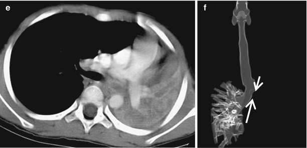
Fig. 14.2
Endobronchial inflammatory myofibroblastic tumor in a 4-year-old boy with cough and fever (Case courtesy of Hye-Kyung Yoon, MD). (a) Plain radiograph shows total opacity of the left lung with mediastinal shifting, suggesting atelectasis of the left lung. (b–e) Nonenhanced CT with lung (b) and mediastinal window (c) and contrast-enhanced CT (d, e) show an endobronchial mass (arrows) occluding the left main bronchus with associated left lung collapse consolidation. (f) 3D volume-rendered image shows abrupt cutoff of the left main bronchus (arrows) and non-visualization of aerated left lung
14.3.1.2 Pleuropulmonary Blastoma
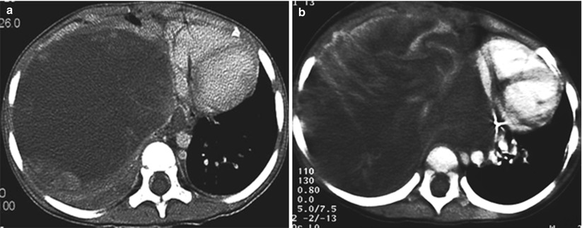
Fig. 14.3
Pleuropulmonary blastoma in a 2-year-old girl. (a, b) Nonenhanced (a) and contrast-enhanced (b) CT show a large, predominantly low-attenuation mass with whorls of high-attenuation soft tissue filling the right thorax and displacing the mediastinal contents
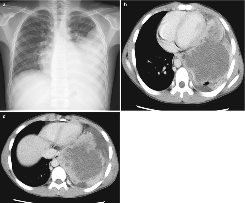
Fig. 14.4
Pleuropulmonary blastoma in a 14-year-old boy. (a) Plain radiograph shows a large opacity in the left hemithorax. (b, c) Contrast-enhanced CT shows mixed cystic and solid masses in the lung periphery adjacent to the pleura in the mid and lower zones. The solid components of the mass lesion and internal septa show enhancement
14.3.1.3 Pulmonary Metastasis
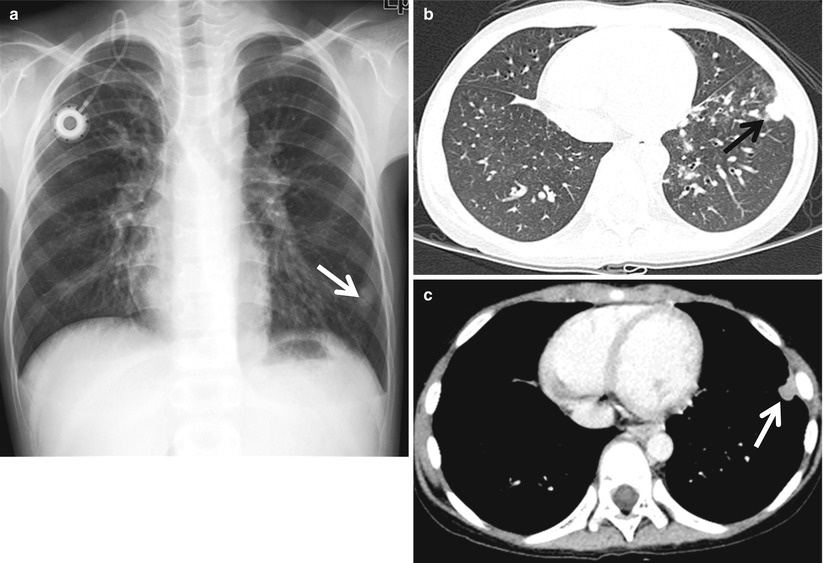
Fig. 14.5
Solitary pulmonary metastasis in a 13-year-old boy with recurrent Wilms tumor. (a) Plain radiograph shows a 15 mm nodule (arrow) in the periphery of the left lower lobe. (b, c) CT with lung window (b) and mediastinal window (c) shows a subpleural-enhancing nodule at the left lower lobe
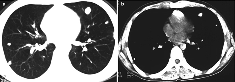
Fig. 14.6
Pulmonary metastases in a 14-year-old boy with known osteosarcoma. (a, b) Nonenhanced CT with lung (a) and mediastinal window (b) shows multiple well-defined calcified nodules, usually occurring in the peripheral areas of the lower lobes
14.3.2 Tumors of the Mediastinum
14.3.2.1 Anterior Mediastinal Tumor
14.3.2.1.1 Lymphoma
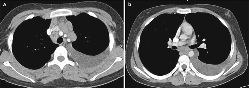
Fig. 14.7
Hodgkin’s disease in a 13-year-old boy with fever and weight loss. (a, b) Contrast-enhanced CT shows discretely enlarged anterior and right paratracheal nodes, lobular anterior and middle mediastinal lymphadenopathy, and left pleural effusion. Lymphomatous nodes appear as homogenous soft tissue attenuation with mild enhancement
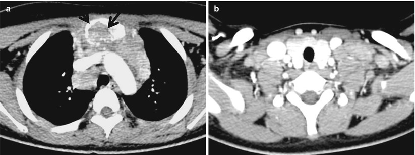
Fig. 14.8
Nodular sclerosing Hodgkin’s disease in a 13-year-old girl with cervical lymphadenopathy. (a) Contrast-enhanced CT at the level of the aortic arch shows right paratracheal lymphadenopathy with a lobular anterior mediastinal mass. Invasion of anterior chest wall and destruction of sternum (arrows) adjacent to the mass are noted. (b) Contrast-enhanced CT at the lower neck shows associated discretely enlarged cervical lymphadenopathy (b)
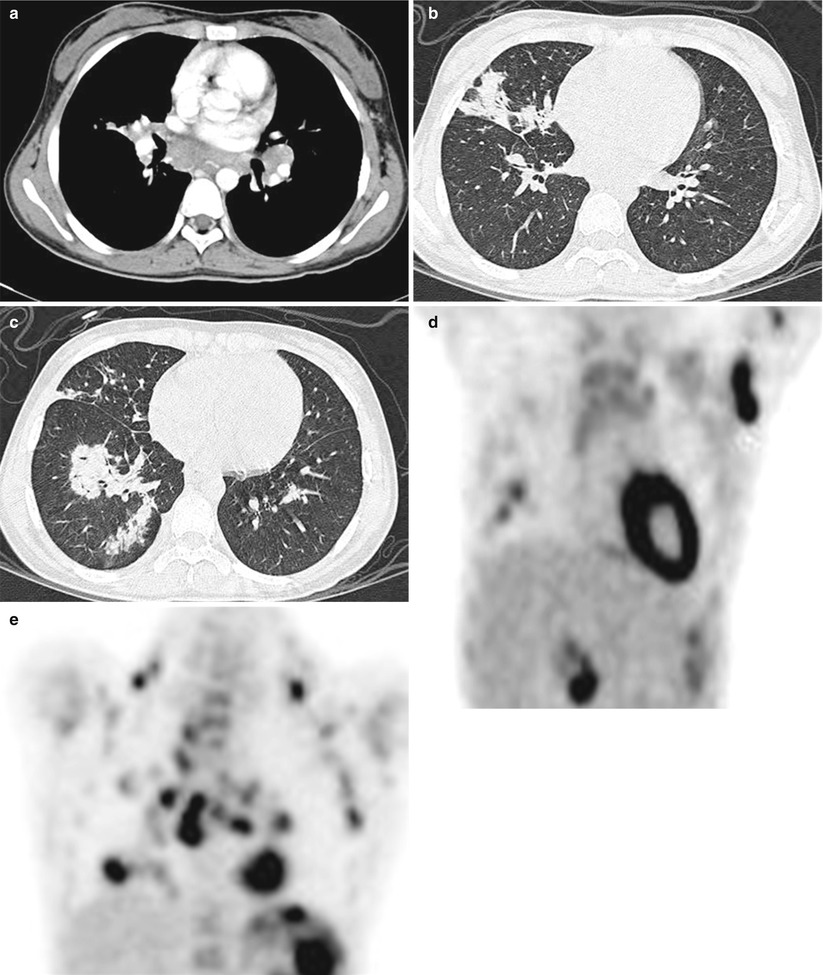
Fig. 14.9
Nodular sclerosing Hodgkin’s disease in a 12-year-old girl with cervical lymphadenopathy. (a) Contrast-enhanced CT with mediastinal window shows bilateral hilar and mediastinal adenopathy with homogenous attenuation. (b, c) CT scans with lung window show confluence and irregular margins of the pulmonary consolidations. (d, e) Coronal FDG-PET demonstrates multiple intense uptakes in the corresponding lymph nodes and lung lesions, seen on CT scans
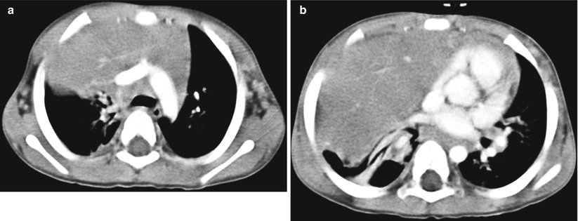
Fig. 14.10
Non-Hodgkin’s lymphoma in a 2-year-old boy with productive cough. (a, b) Contrast-enhanced CT shows an enlarged right thymic lobe. Small linear foci of enhancement represent thymic vessels. Note that enlarged lymph nodes are seen in the paratracheal and subcarinal regions. Note the mass effect of this mass on the adjacent mediastinal structures as well as right lower lobe atelectasis
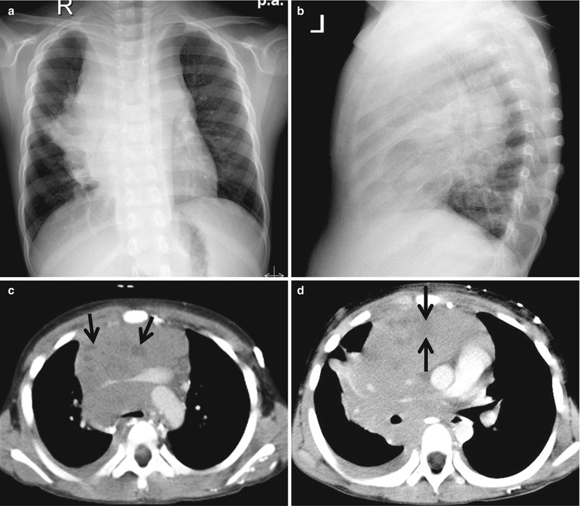
Fig. 14.11
Non-Hodgkin’s lymphoma in a 7-year-old boy with dyspnea. (a) Fontal chest radiograph shows mediastinal widening. (b) Lateral chest radiograph shows that the mass is located in the anterior and middle mediastinum. (c, d) Contrast-enhanced CT shows a lobulated mediastinal mass with the epicenter in the anterior mediastinum. This mass contains some low-attenuation areas of necrosis (arrows). Note the mass effect on the adjacent superior vena cava and enhancing pulmonary vessels. Associated right pleural effusion is noted
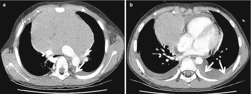
Fig. 14.12
Non-Hodgkin’s lymphoma in a 10-year-old boy with mild fever. (a, b) Contrast-enhanced CT shows a large smooth, biconvex anterior mediastinal mass of uniform attenuation. The trachea and superior vena cava are compressed and displaced, posteriorly. There are also bilateral pleural effusions with pleural masses (arrows) and pericardial effusion
14.3.2.1.2 Germ Cell Tumor: Teratoma
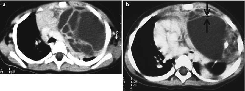
Fig. 14.13
Mature teratoma in a 13-month-old girl with respiratory distress. (a, b) Contrast-enhanced CT shows a large, complex anterior mediastinal mass containing areas of fluid, fat, (arrows) and calcification
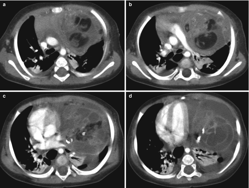
Fig. 14.14


Immature teratoma in a 2-year-old girl with respiratory distress (Case courtesy of Hye-Kyung Yoon, MD). (a–d) Contrast-enhanced CT shows a large, anterior mediastinal mass with variable attenuation with dense calcifications. The mass contains solid and fatty tissues and cystic areas. This complex cystic and fatty immature teratoma displaces the mediastinum to the right
Related posts:


Stay updated, free articles. Join our Telegram channel
Full access? Get Clinical Tree
