Presentation and Presenting Images
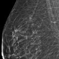
( ▶ Fig. 47.1, ▶ Fig. 47.2, ▶ Fig. 47.3, ▶ Fig. 47.4)
A 55-year-old female presents for screening mammography.
47.2 Key Images
( ▶ Fig. 47.5, ▶ Fig. 47.6, ▶ Fig. 47.7, ▶ Fig. 47.8)
47.2.1 Breast Tissue Density
There are scattered areas of fibroglandular density.
47.2.2 Imaging Findings
Two areas of possible architectural distortion are seen within the left breast. A more subtle area is present in the upper outer quadrant around the 2 o’clock position and the second area is central to the nipple. These are best seen on the digital breast tomosynthesis (DBT) images ( ▶ Fig. 47.5, ▶ Fig. 47.6, ▶ Fig. 47.7, and ▶ Fig. 47.8). The patient had a left breast biopsy 2 years prior, which is marked with a ribbon biopsy clip in the upper inner quadrant. The result of that biopsy was a diagnosis of fibrocystic changes but with findings also suggestive of a radial scar and the usual ductal hyperplasia. The patient elected to observe rather than excise.
47.3 BI-RADS Classification and Action
Category 0: Mammography: Incomplete. Need additional imaging evaluation and/or prior mammograms for comparison.
47.4 Diagnostic Images
( ▶ Fig. 47.9, ▶ Fig. 47.10, ▶ Fig. 47.11, ▶ Fig. 47.12, ▶ Fig. 47.13, ▶ Fig. 47.14, ▶ Fig. 47.15, ▶ Fig. 47.16, ▶ Fig. 47.17)
47.4.1 Imaging Findings
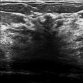
The architectural distortion at 2 o’clock did not persist on further diagnostic imaging (not shown) and the corresponding ultrasound of the area revealed normal breast tissue (not shown). The second area of architectural distortion at 12 o’clock in the middle depth measured approximately 1.9 × 1.3 cm. This area is appreciated on the spot-compression images ( ▶ Fig. 47.9, ▶ Fig. 47.10, and ▶ Fig. 47.11) and on the lateral view ( ▶ Fig. 47.12). The targeted ultrasound revealed an 1.8 × 0.8 × 0.9 cm irregular hypoechoic mass with indistinct and angular margins at 12 o’clock location, 3 cm from the nipple ( ▶ Fig. 47.13 and ▶ Fig. 47.14). The posterior shadowing present helped to define this mass on ultrasound. Additionally, the axillary ultrasound revealed a lymph node 12 cm from the nipple with mild cortical thickening of 4 mm ( ▶ Fig. 47.15). Otherwise all axillary lymph nodes were normal.
47.5 BI-RADS Classification and Action
Category 4C: High suspicion for malignancy
47.6 Differential Diagnosis
Normal breast tissue and breast cancer (invasive ductal carcinoma with metastatic lymph node): More than one lesion can be seen on a screening mammogram. In this case, one was proven to be normal breast tissue and the other cancer.
Radial scar: The imaging features of a radial scar and carcinoma overlap. It is possible that both lesions could be radial scars since the patient has a history of a prior radial scars. The abnormal lymph node does not support a radial-scar diagnosis.
Summation artifact: One “lesion” in question did not persist on DBT and likely represents a summation artifact; however, the second lesion is suspicious and warrants tissue sampling.
47.7 Essential Facts
Architectural distortion accounts for around 6% of imaging detected on screening mammography.
Architectural distortion accounts for 12 to 45% of missed breast cancers.
Architectural distortion is better visualized on DBT compared to full-field digital mammography (FFDM).
In a study by Partyka and colleagues (2014) they found that the cancer detection rate was 21% for architectural distortion lesions seen only on DBT (occult on FFDM).
Architectural distortion has a positive predictive value of a malignancy of nearly 75%. Thus its presence is highly predictive of cancer.
Architectural distortion seen at screening is less likely to represent malignancy compared to architectural distortion seen on a diagnostic mammogram.
47.8 Management and Digital Breast Tomosynthesis Principles
Although architectural distortion is not a very common imaging finding, its detection should warrant complete evaluation due to the high incidence of associated malignancy.
Architectural distortion seen on DBT that does not persist on FFDM and spot compression images, warrants an ultrasound.
Architectural distortion without an ultrasound correlate are more likely to represent a radial sclerosing lesion than carcinoma. Larger studies are needed to determine if lesions falling into this imaging situation can be watched or need to be biopsied.
47.9 Further Reading
[1] Bahl M, Baker JA, Kinsey EN, Ghate SV. Architectural Distortion on Mammography: Correlation With Pathologic Outcomes and Predictors of Malignancy. AJR Am J Roentgenol. 2015; 205(6): 1339‐1345 PubMed
[2] Partyka L, Lourenco AP, Mainiero MB. Detection of mammographically occult architectural distortion on digital breast tomosynthesis screening: initial clinical experience. AJR Am J Roentgenol. 2014; 203(1): 216‐222 PubMed

Fig. 47.1 Left craniocaudal (LCC) mammogram.
Stay updated, free articles. Join our Telegram channel

Full access? Get Clinical Tree


