and Zdeněk Fryšák1
(1)
Department of Internal Medicine III – Nephrology, Rheumatology and Endocrinology, Faculty of Medicine and Dentistry, Palacky University Olomouc and University Hospital Olomouc, Olomouc, Czech Republic
Keywords
Nodular goiterUltrasoundFine-needle aspiration biopsyThe Bethesda Cytologic Classification24.1 Essential Facts
Diagnosis of thyroid nodules by needle biopsy was first described by Martin and Ellis in 1930; they used an 18-gauge-needle aspiration technique. Scandinavian investigators introduced “small-needle” aspiration biopsy of the thyroid in the 1960s, and this technique came into widespread use in North America in the 1980s [1, 2].
24.2 Importance and Benefits of FNAB in the Diagnosis of Nodular Thyroid Disease in the Beginning of the 1990s by Gharib [4]
Efficacy of Fine-Needle Aspiration Biopsy (FNAB) and its role in the management of a nodular goiter was clearly established. Accuracy of cytologic diagnosis approached 95%.
FNAB is a reasonable approach for thyroid nodules; its costs have decreased substantially because it facilitates selection of patients needing to undergo surgical excision.
Selecting patients for operation on the basis of results of FNAB has more than doubled the yield of carcinoma.
Limitations of cytologic examination, nondiagnostic results, and cellular follicular neoplasms should be remembered but need not negate continued use of FNAB.
Negative (benign) and positive (malignant) cytologic results are conclusive; careful clinical follow-up of benign nodules and surgical excision of malignant nodules are recommended.
Nondiagnostic results are inconclusive; further evaluation by repeated FNAB, ultrasound-guided biopsy, or radionuclide scanning is necessary.
Suspicious cytologic results are also inconclusive and are associated with a 20% chance of malignant involvement; surgical treatment is necessary for clarification.
FNAB is a safe, inexpensive, minimally invasive, highly accurate, and cost-effective means in the diagnosis of nodular thyroid disease.
The procedure has a central role in the management of thyroid nodules and should be used as the initial diagnostic test.
The introduction of FNAB had a substantial effect on the management of patients with thyroid nodules. The percentage of patients undergoing thyroidectomy decreased by 25%, and the yield of carcinoma in patients who undergo surgery increased from 15% to at least 30%. FNAB decreased the cost of care by 25% [5].
24.3 Diagnostic Yield of FNAB in the Diagnosis of Nodular Thyroid Disease in the Beginning of the 1990s by Gharib [5]
Four cytologic diagnostic categories are used. Prevalence of these categories: benign, 69%; suspicious, 10%; malignant, 4%; and nondiagnostic, 17%.
Analysis of FNAB data suggests false-negative rate of 1–11%, false-positive rate of 1–8%, sensitivity of 65–98%, and specificity of 72–100%.
Limitations of FNAB are related to the skills of the aspirator, the expertise of the cytologist, and the difficulty in distinguishing some benign cellular adenomas from their malignant counterparts.
24.4 Current FNAB Experience by Dean and Gharib [6]
Thyroid FNAB is the most accurate test to determine malignancy, and is an integral part of current thyroid nodule evaluation. Results are superior when US-guided FNAB is performed.
FNAB results are classified as diagnostic (satisfactory) or nondiagnostic (unsatisfactory). Unsatisfactory smears (5–10%) result from hypocellular specimens usually caused by cystic fluid, bloody smears, or suboptimal preparation.
Diagnostic smears are conventionally subclassified into benign, indeterminate, or malignant categories:
Benign cytology (60–70%) is negative for malignancy, and includes cysts, colloid nodule, or Hashimoto’s thyroiditis.
Malignant cytology (5%) is almost always positive for malignancy, and includes primary thyroid tumors or nonthyroid metastatic cancers.
Indeterminate or suspicious specimens (10–20%) include atypical changes, Hurthle cells, or follicular neoplasms.
The new Bethesda Cytologic Classification has a six-category classification, further subdividing indeterminate by risk factors. Overall, the indeterminate category has anywhere from 15% up to 60% risk of malignancy.
Recent development of molecular markers should help to further separate benign from malignant nodules with an indeterminate cytology.
Caution! Benign lesions on FNAB have an approximate 3% risk of malignancy, and may be followed with US. Repeating FNAB decreases the risk of a false negative result to 1.3% [7].
24.5 The Large Analysis of the Brigham and Women’s Hospital and Harvard Medical School with a Total of 1985 Patients and FNAB of 3483 Nodules [8]
Typical malignant nodule: solid (14.3%), hypoechoic (15.2%), with punctuate calcifications (23.3%), solitary (14%).
Prevalence of thyroid cancer was similar between the patients with solitary nodule (14.8%) and the patients with multiple nodules (4.9%).
Solitary nodules represent twice the risk of malignancy when compared to nonsolitary nodules and more than 1.5 times higher in a man than in a woman.
In patients with multiple nodules ≥10 mm, cancer was multifocal in 46%, and 72% of cancers occurred in the largest nodule.
Risk ranges from a 48% likelihood of malignancy in a solitary solid nodule with punctate calcifications in a man to less than 3% in a noncalcified predominantly cystic nodule in a woman.
In patients with one or more thyroid nodules ≥10 mm, likelihood of thyroid cancer per a patient is independent of number of nodules, whereas likelihood per nodule decreases with increasing number of nodules.
In patients with two or more nodules, aspiration of only the largest nodule would have missed almost one-third of the malignancies. For exclusion of cancer in a thyroid with multiple nodules ≥10 mm, up to four nodules should be considered for FNAB. Sonographic characteristics can be used to prioritize nodules for FNAB based on their individual risk of cancer.
24.6 Recommendations for Diagnostic FNAB of a Thyroid Nodules Based on US Pattern According to the 2015 ATA Guidelines [9]
Features with the highest specificity (median > 90%) for thyroid cancer are microcalcifications, irregular margins, and taller-than-wide shape.
Diagnostic FNAB of a nodule <10 mm should be performed only in suspicious cases or in patients with personal history of radiation exposure or familial thyroid cancer.
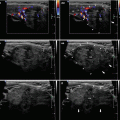
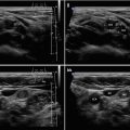
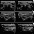
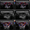
US patterns and FNAB guidance for thyroid nodules: see Table 24.1.
Table 24.1
Sonographic patterns, estimated risk of malignancy, and US-FNAB guidance for thyroid nodules
Sonographic pattern | US features | Estimated risk of malignancy, % | FNA size cutoff (largest dimension) |
|---|---|---|---|
High suspicion | Solid hypoechoic nodule or solid hypoechoic component of a partially cystic nodule with one or more of the following features: irregular margins (infiltrative, microlobulated), microcalcifications, taller-than-wide shape, rim calcifications with small extrusive soft tissue component, evidence of ETE | >70–90 | Recommend FNAB at ≥1 cm |
Intermediate suspicion
Stay updated, free articles. Join our Telegram channel
Full access? Get Clinical Tree
 Get Clinical Tree app for offline access

|
