Chapter 42 Ultrasound imaging
Chapter contents
42.1 Aim
The aim of this chapter is to discuss the key properties of sound and explain how sound can be used to produce a diagnostic image. The Doppler principle, by which the velocity of moving tissues can be determined, is introduced. The clinical applications and advantages of ultrasound are described.
42.2 Sound properties
The speed and amplitude of sound is greatly affected by the medium through which it passes, much more so than in the case of electromagnetic radiations such as light. You may have noticed the very different pitches of sound that you hear when your head is under water, compared with when your head breaks back above the surface. Sound travels at slightly different speeds in different body tissues. The speed of sound is affected by the compressibility of the medium. It travels faster in more rigid materials which resist being compressed, and more slowly in materials such as fluids and gases which can be compressed easily. As an analogy, think of how much easier you can push an object using a firm rod made of wood, compared with using a bendy rod made of rubber. Table 42.1 lists the speed of sound in air and biological tissues and it can be seen that the more rigid substances allow sound to travel faster. Reflection and refraction of sound can occur at boundaries between materials or tissues.
Table 42.1 Approximate speeds of sound in different materials and tissues
| MATERIAL OR TISSUE | SPEED IN METRES PER SECOND |
|---|---|
| Air at 20°C and normal atmospheric pressure | 340 |
| Lung | 650 |
| Fat | 1460 |
| Pure water | 1500 |
| Salt water | 1530 |
| Kidney | 1560 |
| Blood | 1570 |
| Muscle | 1580 |
| Bone | 3000 |
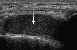
Reflection is a very important property of sound waves, as it provides echoes at tissue boundaries, and these are used to depict structures in diagnostic ultrasound. It also gives some information about the nature of tissues. A cyst, which mostly contains fluid, will return few or no echoes (the signal is termed hypoechoic or anechoic) from within itself, as shown in Figure 42.1, while a haemorrhage will return echoes from within itself (the signal is echoic or hyperechoic), as shown in Figure 42.2. This is because a haemorrhage also contains blood cells and proteins (which return echoes) in addition to just fluid. The walls of a cyst may return strong echoes at the capsule–fluid boundary.
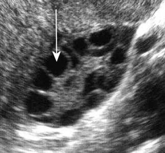
Figure 42.1 The cysts within this polycystic ovary contain watery fluid and appear anechoic (echo free).
• Soft tissue/air interface – large Z difference=large reflection.
• Soft tissue/bone interface – large Z difference=large reflection (about 100%).
The acoustic impedances of body tissues can be seen in Table 42.2.
Table 42.2 Acoustic impedance (Z) values for air and body tissues
| MATERIAL OR TISSUE | ACOUSTIC IMPEDANCE (KG.M−2.S−1) |
|---|---|
| Air | 0.004 × 10⁶ |
| Fat | 1.34 × 10⁶ |
| Water | 1.48 × 10⁶ |
| Liver | 1.65 × 10⁶ |
| Blood | 1.65 × 10⁶ |
| Muscle | 1.71 × 10⁶ |
| Bone | 7.8 × 10⁶ |
Large Z differences return good strong echoes but
Stay updated, free articles. Join our Telegram channel

Full access? Get Clinical Tree


