Abstract
Unilateral renal agenesis has a birth prevalence of approximately 1 : 1000 to 1 : 2000, but it is often undetected prenatally. Sonographically, one kidney cannot be visualized in either the renal fossa or the pelvis, and the adrenal flattens and fills the renal fossa—the “lying-down” adrenal sign. Color Doppler imaging of the descending aorta reveals just one renal artery. The solitary kidney often develops compensatory hypertrophy. Unilateral renal agenesis is associated with other anomalies, particularly genitourinary abnormalities, and it is a component of several genetic syndromes. Therefore, targeted sonography is indicated, and amniocentesis may be offered. Affected individuals are usually asymptomatic; however, they are at increased risk to develop hypertension and renal insufficiency.
Keywords
renal fossa, lying-down adrenal sign, solitary kidney
Introduction
Unilateral renal agenesis, also called solitary kidney, is a common congenital anomaly. Affected individuals are usually asymptomatic, and if a diagnosis is not made prenatally, they may not receive medical attention until complications arise. If unilateral renal agenesis is diagnosed in childhood, nearly 50% of affected children have an associated renal abnormality, including 30% with vesicoureteral reflux (VUR). Being born with one kidney is not equivalent to reaching adulthood with two kidneys and subsequently undergoing a nephrectomy. Individuals with unilateral renal agenesis, even in the absence of reflux, have an increased risk of hypertension, renal insufficiency, and progression to end-stage renal disease requiring transplant. Earlier identification of unilateral renal agenesis may forestall the development of these complications. Improvements in ultrasound (US) technology have made it easier to identify unilateral renal agenesis with prenatal US.
Disorder
Definition
Unilateral renal agenesis is the congenital absence of one kidney and ureter.
Prevalence and Epidemiology
Unilateral renal agenesis occurs in approximately 1 : 1000 to 1 : 2000 births, but it often goes undetected prenatally. In a systematic review, unilateral renal agenesis was diagnosed prenatally in 1 : 8000 pregnancies, and in a population-based registry of European countries with routine sonography, unilateral renal agenesis was identified prenatally in only 1 : 20,000 pregnancies. This is in contrast to the excellent detection of bilateral renal agenesis, and it highlights the importance of maintaining an index of suspicion. Male predominance of unilateral renal agenesis has been reported in some studies but not others. The left kidney is absent more often than the right. Unilateral renal agenesis is also more common in the setting of pregestational diabetes.
Associated renal anomalies, including VUR, ureteropelvic junction (UPJ) obstruction, and ureterovesical junction obstruction, have been reported in 30% to 50% of children referred for evaluation. In females, müllerian anomalies are common, particularly bicornuate uterus, uterus didelphys, and unicornuate uterus. Occasionally, these associated anomalies are first identified in the setting of Herlyn-Werner-Wunderlich syndrome (uterus didelphys, obstructed hemivagina, and ipsilateral renal agenesis) because of hematocolpos at the time of initiation of menstruation during puberty. In males, there is an increased incidence of congenital absence of the vas deferens and cysts of the seminal vesicles. Other associations include caudal dysgenesis, skeletal anomalies, and malformations of the central nervous system. Unilateral renal agenesis is a component of several syndromes, including branchiootorenal syndrome, 22q11.2 deletion syndrome (see Chapter 154 ), and Fraser syndrome (see Chapter 128 ), and it is one of several renal anomalies encountered in the VACTERL ( v ertebral, a nal, c ardiac, t racheal, e sophageal, r enal, and l imb anomalies) association (see Chapter 146 ). The aneuploidy risk is increased in the setting of other anomalies.
Etiology and Pathophysiology
Renal agenesis results when the ureteric bud either fails to develop from the mesonephric (Wolffian) duct or fails to induce the surrounding metanephric mesenchyme to form glomeruli and nephrons. There is a significant familial aggregation of cases. Offspring of individuals with unilateral renal agenesis have a 12% incidence of urogenital anomalies, more than 50% of which are renal agenesis; there is also an increased risk among other first-degree relatives. In one series, 20% of cases of unilateral renal agenesis had evidence of a mutation in the RET (Rearranged in Transfection) gene.
Individuals with unilateral renal agenesis are at increased risk of progression to end-stage renal disease, postulated to be caused by an overall reduction in their number of nephrons, with subsequent hyperfiltration and focal glomerulosclerosis. From the standpoint of prenatal or postnatal imaging, it may be impossible to differentiate true renal agenesis from severe hypoplasia (see Chapter 9 ) or multicystic dysplasia with subsequent regression (see Chapter 15 ), and individuals with these lesions may have abnormalities of the contralateral, normal-appearing kidney as well.
Manifestations of Disease
Clinical Presentation
The prenatal diagnosis of unilateral renal agenesis is based on US findings, as discussed subsequently. This anomaly commonly goes undetected prenatally and postnatally. Children may be diagnosed during evaluation for urinary tract infection because of the association with VUR. Adults may present with hypertension, proteinuria, or renal insufficiency. Unilateral renal agenesis may remain undiagnosed in many individuals throughout life.
Imaging Technique and Findings
Ultrasound.
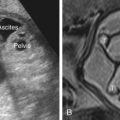
Typically, the initial US finding is an inability to visualize a normal kidney in the renal fossa ( Fig. 11.1 ). When the diagnosis is suspected, it is important to image the renal fossae in multiple planes and to look for a pelvic kidney (see Chapter 8 ). If a kidney is not present in the renal fossa, the adrenal flattens and fills the fossa in what has been termed the “lying-down” adrenal sign ( Fig. 11.2 ). The adrenal gland is suprarenal and almond-shaped, with a hypoechoic cortex and hyperechoic medulla.


Color Doppler imaging of the descending aorta may be a useful adjunct. The fetal abdomen is imaged in the coronal plane with the beam perpendicular to the aorta so that renal arteries arise at an angle of insonation below 20 degrees. A renal artery is visible only on the side that has a kidney ( Fig. 11.3 ).