A. Tuberous sclerosis
B. Diabetes mellitus
C. Hypertension
D. Urothelial carcinoma
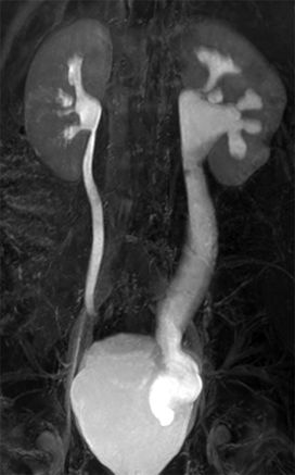
5 A 3-year-old boy with a congenital left ureteropelvic junction (UPJ) obstruction related to a crossing vessel undergoes MR urography. The image on the left was acquired with a repetition time (TR) of 2,000 msec and an echo time (TE) of 200 msec. The image on the right was obtained at the same level with a TR of 3.6 msec and a TE of 1.8 msec. What is the most likely cause of the hypointense material in the left renal pelvis on the right-hand image?
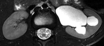
A. Hemorrhage
B. Malignancy
C. Contrast material
D. Parasitic infection
E. The etiology cannot be determined with the provided images.
6 A 25-year-old male with vesicoureteric reflux presents with right flank discomfort, and an ultrasound is obtained. The arcuate arteries are sampled within each kidney and compared to assess for symmetry. What is the formula for calculating the resistive index?

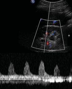
A. (Peak systolic velocity − End-diastolic velocity)/Peak systolic velocity
B. (End-diastolic velocity − Peak systolic velocity)/Peak diastolic velocity
C. (Peak systolic velocity − End-diastolic velocity)/End-systolic velocity
D. (End-diastolic velocity − Peak systolic velocity)/End-diastolic velocity
E. End-diastolic velocity/Peak systolic velocity
F. End-systolic velocity/Peak diastolic velocity
G. End-diastolic velocity/Peak diastolic velocity
H. End systolic velocity/Peak systolic velocity
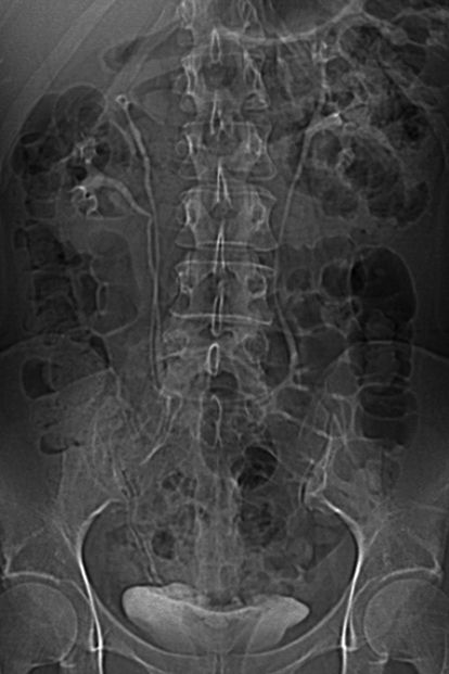
7 A 62-year-old male with a 40-pack-year smoking history, chronic obstructive pulmonary disease, and hematuria presents for a CT urogram and subsequent right retrograde pyelogram. What are the two most common causes of this imaging finding?
A. Urothelial cancer and Mycobacterium tuberculosis
B. Urothelial cancer and renal calculus disease
C. Schistosoma haematobium and Proteus mirabilis
D. Escherichia coli and Proteus mirabilis
8 A 22-year-old pregnant female in the third trimester with suspected stone disease undergoes an ultrasound examination that demonstrates moderate dilation of the proximal right collecting system and asymmetric resistive indices. The referring service requests a low-dose unenhanced renal stone protocol CT of the abdomen and pelvis. If the effective dose to the fetus is 10 mSv, what is the approximate risk of radiation-induced cancer conferred to the fetus?
A. The exact risk is speculative, but it is estimated to be roughly 1 in 250 (0.4%).
B. The exact risk is speculative, but it is estimated to be roughly 1 in 25 (4%).
C. The exact risk is speculative, but it is estimated to be roughly 1 in 5 (20%).
D. The exact risk is speculative, but it is estimated to be roughly 1 in 2 (50%).
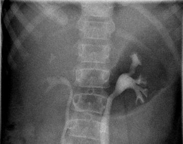
9 A 42-year-old female with microhematuria undergoes an intravenous urogram. The only identifiable abnormality is a completely duplicated right collecting system (shown below). With respect to this finding, what does the Weigert-Meyer rule predict?
A. The upper pole moiety will insert superior and medial to the lower pole moiety.
B. The upper pole moiety will insert inferior and medial to the lower pole moiety.
C. The upper pole moiety will insert superior and lateral to the lower pole moiety.
D. The upper pole moiety will insert inferior and lateral to the lower pole moiety.
10 A 22-year-old nonsmoking female with no past medical history presents with asymptomatic microhematuria (≥3 red blood cells per high-powered field) detected during a preemployment urinalysis. A triphasic CT urogram is ordered. What is the best next step?
A. Perform the CT urogram as ordered.
B. Recommend a limited CT urogram (two of three phases).
C. Recommend an unenhanced CT.
D. Consider cancelling the test.
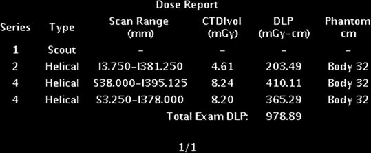
11 A 40-year-old male with recurrent urolithiasis undergoes an unenhanced CT of the abdomen and pelvis. The following exam card is reported by the scanner at the end of the study. What does the number 15.42 represent?
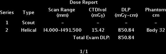
A. It is the radiation dose delivered to the patient.
B. It is the scanner-specific radiation output.
C. It is the radiation dose delivered to a hypothetical patient.
D. It is the radiation output delivered by a hypothetical scanner.
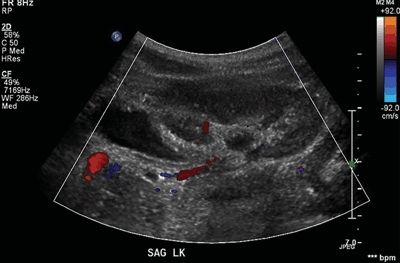
12 A 32-year-old pregnant female in her second trimester presents with right flank pain. A retroperitoneal ultrasound is performed, demonstrating bilateral ureteral jets. Which of the following best explains the principle of color Doppler imaging?
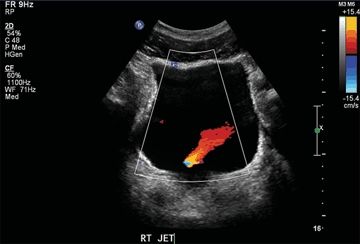
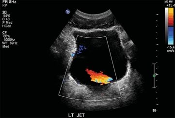
A. The reflected ultrasound frequency always increases when the ultrasound beam interacts with a structure moving toward it.
B. The reflected ultrasound wavelength always increases when the ultrasound beam interacts with a structure moving toward it.
C. Red pixels are always assigned when the ultrasound beam interacts with a structure moving toward it.
D. Blue pixels are always assigned when the ultrasound beam interacts with a structure moving toward it.
13 A 55-year-old male with gross hematuria undergoes a CT urogram consisting of unenhanced, nephrographic, and excretory phase images of the abdomen and pelvis. The following exam card is reported by the scanner at the end of the study. What is the effective dose to the patient?
A. 8.24 mGy
B. 21.05 mGy
C. 978.89 mGy-cm
D. The effective dose cannot be determined with these data.
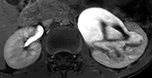
14 A 5-year-old boy with congenital orthotopic megaureter undergoes MR urography to assess anatomy and function. Three-dimensional reconstructions were performed of the coronal source data. The image on the left was obtained with a repetition time (TR) of 4,000 msec and an echo time (TE) of 200 msec. The image on the right was obtained with a TR of 3.6 msec and a TE of 1.8 msec. What is the approximate relative function of the two kidneys?
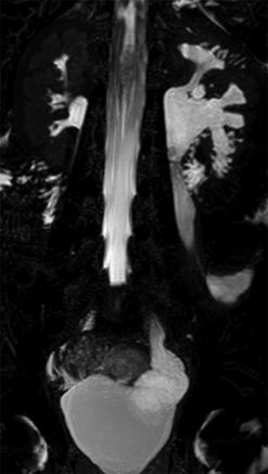
A. The right kidney is substantially less functional than is the left kidney.
B. The left kidney is substantially less functional than is the right kidney.
C. The two kidneys have grossly similar function.
D. The relative function of the kidneys cannot be estimated with these images.
15 A 52-year-old male with diabetes mellitus, fever, and leukocytosis presents with left flank pain. Vital signs are as follows: pulse 115, blood pressure 89/65, respiratory rate 22, SpO2 98% on room air. The following ultrasound is obtained. What is the most likely diagnosis?
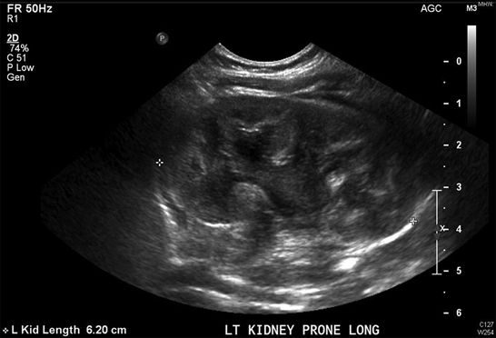
A. Pyelonephritis
B. Pyelitis
C. Pyonephrosis
D. Emphysematous pyelitis
E. Emphysematous pyelonephritis
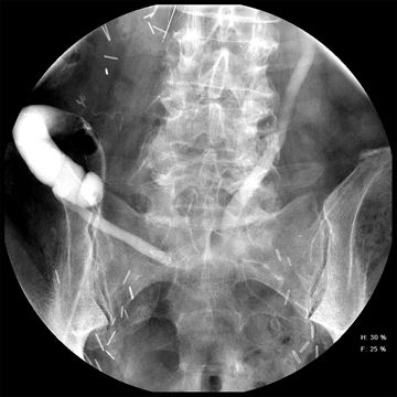
16 A 7-year-old girl with recurrent pyelonephritis undergoes a voiding cystourethrogram (VCUG). What is the diagnosis?
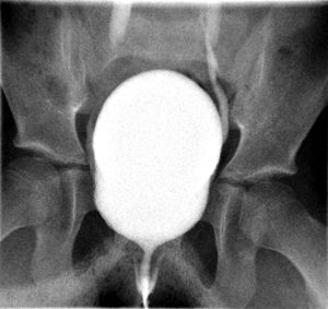
A. Normal study
B. Bilateral grade I vesicoureteric reflux
C. Bilateral grade II vesicoureteric reflux
D. Bilateral grade IV vesicoureteric reflux
17 A 70-year-old male undergoes bilateral percutaneous nephrostomy catheter placement. Which of the following best characterizes the imaging findings in the midportions of the ureters?
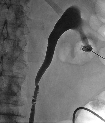
A. Malignant condition associated with urothelial carcinoma
B. Benign condition associated with urothelial carcinoma
C. Benign vascular impressions
D. Benign ureteral “kinks”
18 An 8-year-old girl with recurrent abdominal pain undergoes a retroperitoneal ultrasound. Which of the following is the best explanation for the imaging finding(s) in the left kidney?
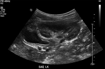
A. Multicystic dysplastic kidney
B. Autosomal dominant polycystic kidney disease
C. Acquired renal cyst
D. Obstructed ectopic ureter
E. Autosomal recessive polycystic kidney disease
19 A 55-year-old male with asymptomatic microscopic hematuria undergoes a CT urogram. Representative images below depict an abnormality in the left kidney. What is the most appropriate management for this entity?
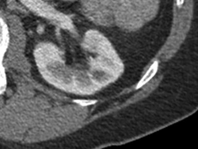
A. Ignore (benign finding)
B. Prophylactic antibiotics
C. Percutaneous aspiration
D. Embolization
E. Operative resection
20 A 50-year-old male with recurrent urolithiasis undergoes an unenhanced CT of the abdomen and pelvis with a CTDIvol of 14 mGy based on a 32-cm-diameter body dosimetry phantom. His effective diameter is 17 cm, which translates to a 1.98
 conversion factor, and the DLP is 700 mGy-cm (scan length: 50 cm). What is this patient’s size-specific dose estimate (SSDE)?
A. 7.1 mGy
B. 27.7 mGy
C. 353.5 mGy-cm
D. 1,386 mGy-cm
E. The SSDE cannot be determined with these data.
21 A 60-year-old male with gross hematuria and a negative cystoscopy undergoes a CT urogram that demonstrates an abnormality in the left proximal collecting system. Which of the following is a correct comparison between upper tract urothelial cancer and bladder cancer?
A. Upper tract urothelial cancer is more common than is bladder cancer.
B. Upper tract urothelial cancer is less likely to be invasive than is bladder cancer.
C. Smoking is a risk factor for bladder cancer but not for upper tract urothelial cancer.
D. Aromatic hydrocarbons cause both bladder cancer and upper tract urothelial cancer.
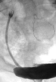
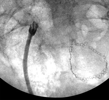
22 A 63-year-old male with a history of urothelial carcinoma of the right renal pelvis and bladder, and who is status post right nephroureterectomy, cystoprostatectomy, and ileal loop creation with an end-to-end anastomosis, undergoes a loopogram to assess for leak. How should the contrast material in the left collecting system be characterized?
A. Abnormal spontaneous reflux, grade I
B. Abnormal spontaneous reflux, grade III
C. Abnormal spontaneous reflux, grade V
D. Expected finding
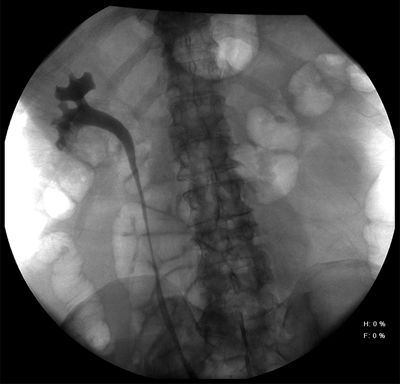
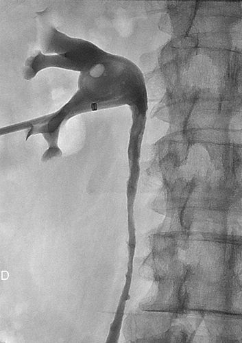
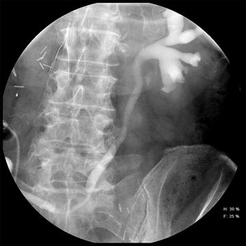
23 A 62-year-old male with hydronephrosis undergoes a right retrograde pyelogram. What is the name of this finding, and what does it imply?
A. Goblet sign, which supports the diagnosis of a urothelial malignancy
B. Goblet sign, which supports the diagnosis of a ureteral calculus
C. Comet tail sign, which supports the diagnosis of a urothelial malignancy
D. Comet tail sign, which supports the diagnosis of a ureteral calculus
24 A 2-year-old girl with an abnormal abdominal ultrasound undergoes a voiding cystourethrogram (VCUG). What is the most likely diagnosis?
A. Grade III reflux
B. Grade III reflux and congenital duplication
C. Opacification of an obstructed collecting system
D. Opacification of a normal collecting system
25 Unnecessary renal stone CT examinations are commonly cited as a major source of radiation exposure in young patients, and efforts are underway to minimize the radiation dose from these examinations. What is the principal effect of statistical iterative reconstruction?
A. Improve image signal
B. Improve image contrast
C. Decrease radiation dose
D. Decrease image noise
26 A 25-year-old male with right lower quadrant pain and suspected appendicitis presents for a contrast-enhanced CT of the abdomen and pelvis. The effective dose is estimated to be 10 mSv. How does this compare to the typical annual natural background radiation dose for United States citizens living at sea level?
A. It is approximately five times the annual background dose of 2 mSv.
B. It is approximately 50 times the annual background dose of 0.2 mSv.
C. It is approximately 500 times the annual background dose of 0.02 mSv.
D. It is approximately 5,000 times the annual background dose of 0.002 mSv.
27 A 32-year-old female with left flank pain and hematuria undergoes an unenhanced renal stone protocol CT of the abdomen and pelvis. Which imaging sign best characterizes the more anterior calcification in the left hemipelvis, and what does it imply?
A. Comet tail sign, which supports the diagnosis of a ureteral calculus
B. Comet tail sign, which supports the diagnosis of a phlebolith
C. Soft-tissue “rim” sign, which supports the diagnosis of a ureteral calculus
D. Soft-tissue “rim” sign, which supports the diagnosis of a phlebolith
28 A 50-year-old male with recurrent urinary tract infections undergoes an abdominal radiograph demonstrating a large calcification in the right upper quadrant. This calcification is likely comprised of what dominant material?
A. Calcium oxalate
B. Calcium phosphate
C. Struvite
D. Cystine
E. Uric acid
29 A radiology practice is interested in updating their abdominal CT scan protocols to reduce the radiation dose to their patients. What effect would a lower kVp setting (e.g., 80 kVp instead of 120 kVp) have on the resultant images?
A. Decreased image noise
B. Increased radiation dose
C. Increased attenuation of iodine
D. Decreased pseudoenhancement
30 A new imaging modality is developed for the detection of upper tract urothelial carcinoma. Its feasibility is tested in a small patient population. The test identifies cancer in three patients with the disease and two patients without the disease. The test is “negative” in one patient with the disease and eight patients without the disease. What are the sensitivity and specificity of this test based on these data?
A. Sensitivity: 75%, specificity: 20%
B. Sensitivity: 25%, specificity: 80%
C. Sensitivity: 75%, specificity: 80%
D. Sensitivity: 25%, specificity: 20%
Answers and Explanations
1 Answer B. Dual-energy CT is based on the principle of distinguishing elements that have sufficiently unique K-edges by imaging them with two different kVp settings. When an atom is struck by a photon of sufficient energy to dislodge an electron from the K-shell of that atom, the electron can be discharged and replaced by an electron from a neighboring ring. When this occurs, an x-ray photon is discharged. This phenomenon is known as the photoelectric effect.
Just above the K-shell binding energy is the K-edge, which is characterized by a sudden increase in attenuation at that energy level caused by a sudden increase in the probability of the photoelectric effect occurring. The K-edge is element specific and increases with increasing atomic number. When two elements with sufficiently unique K-edges are imaged with two different kVp settings (most commonly 80 and 140 kVp on clinical scanners), it is possible to determine the composition of those elements by the way they behave in the radiation environment. The attenuation of those elements at varying kVp settings gives information about their K-edge, and allows one to determine indirectly what they are.
Most elements that constitute the human body (e.g., carbon, hydrogen, oxygen, nitrogen) have very similar K-edges (range: 0.01 to 0.53 keV), making them unlikely candidates for dual-energy separation. However, minerals (e.g., calcium [K-edge: 4.0 keV]) and iodine (e.g., iodinated contrast material [K-edge: 33.2 keV]) are different enough from background tissue that separation is possible. Once separation is achieved, those elements can be characterized, quantified, and/or removed from the image. This forms the foundation of dual-energy characterization of renal calculi and permits the creation of “virtual unenhanced” CT.
References: Boll DT, Patil NA, Paulson EK, et al. Renal stone assessment with dual-energy multidetector CT and advanced postprocessing techniques: improved characterization of renal stone composition—Pilot study. Radiology 2009;250:813–820.
Heye T, Nelson RC, Ho LM, et al. Dual-energy CT applications in the abdomen. AJR Am J Roentgenol 2012;199:S64–S70.
Williams JC Jr, Saw KC, Paterson RF, et al. Variability of renal stone fragility in shock wave lithotripsy. Urology 2003;61:1092–1096.
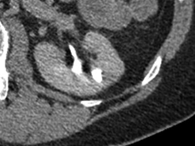
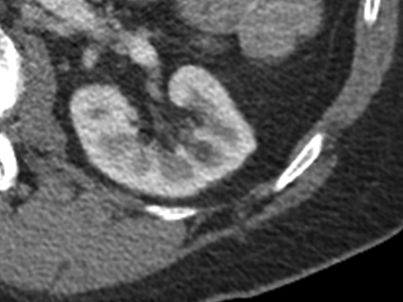
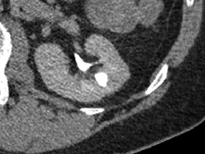
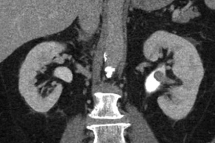
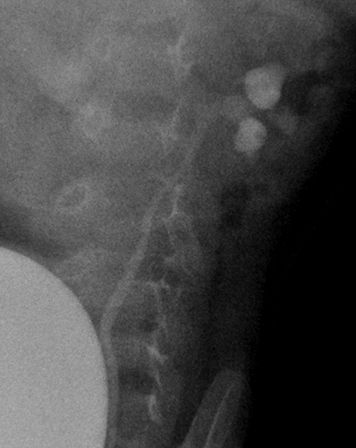
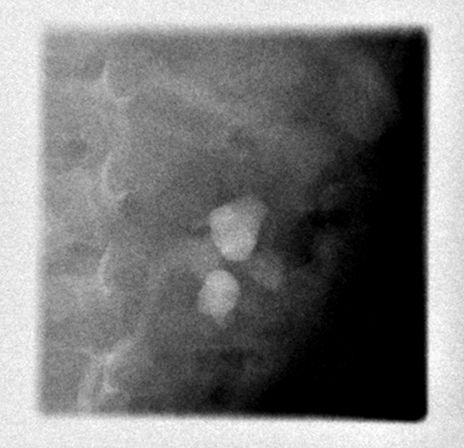
2 Answer D. Medullary sponge kidney is characterized by medullary nephrocalcinosis and renal tubular ectasia. Renal tubular ectasia is characterized by radiating parallel linear contrast arrays at the medullary tips (two examples shown below); it should be distinguished from the normal “papillary blush,” which is homogeneous, vague, and nonlinear. The underlying abnormality in renal tubular ectasia is a defect in the renal tubules that results in tiny sac-like cysts that impair urine transit and predispose to stone formation. Many patients with renal tubular ectasia do not have calculi or systemic signs of renal disease; in these patients, the ectatic ducts are often thought to be an incidental finding.
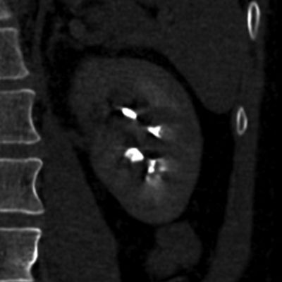
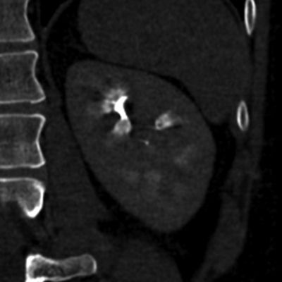
Renal tubular ectasia, characterized by radiating tubular collections at the medullary tips
The differential diagnosis for medullary nephrocalcinosis is:
- Medullary sponge kidney [common cause]
- Type I (distal) renal tubular acidosis [common cause]
- Hyperparathyroidism [common cause]
- Other causes of hypercalcinosis (e.g., hypervitaminosis D, milk–alkali syndrome)
- Sarcoidosis
- Oxaluria
- Furosemide use
The differential diagnosis for cortical nephrocalcinosis is:
- Renal cortical necrosis (secondary to severe systemic hypotension) [common cause]
- Chronic glomerulonephritis [common cause]
- Chronic pyelonephritis
- Alport syndrome
- Oxaluria
Related posts:


Stay updated, free articles. Join our Telegram channel

Full access? Get Clinical Tree


