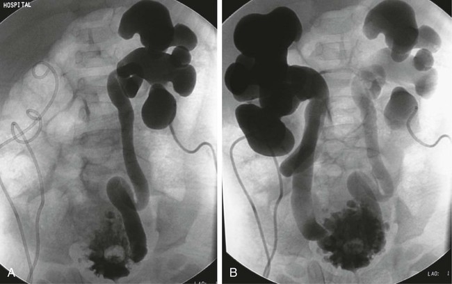
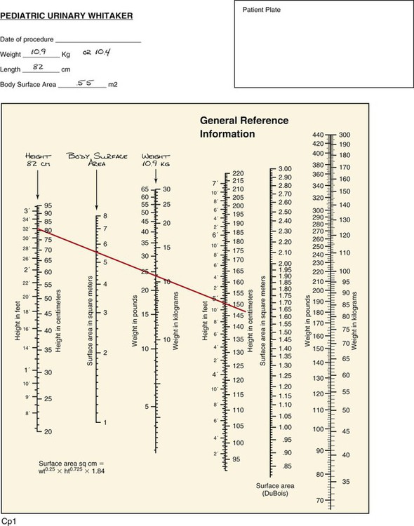
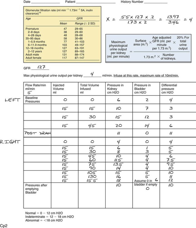
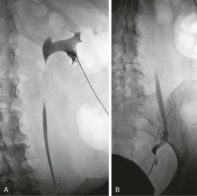
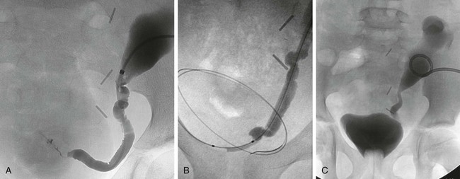
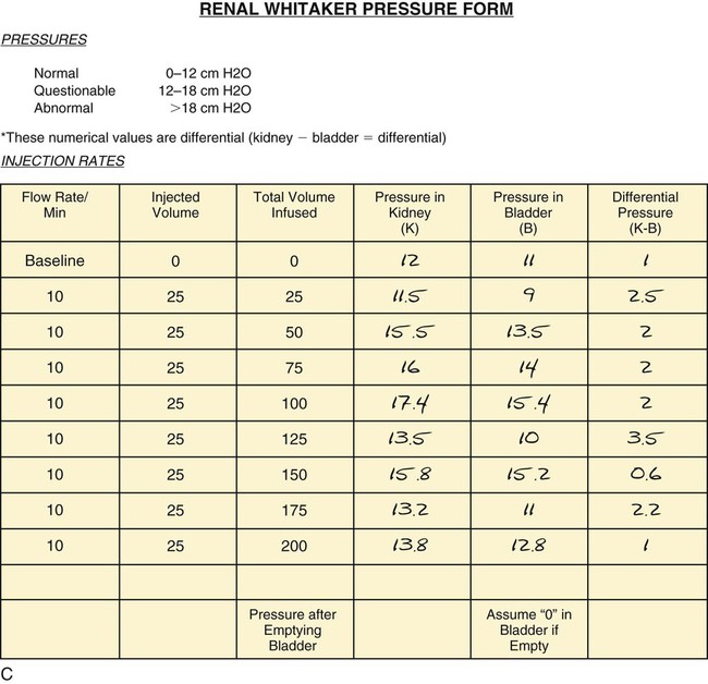
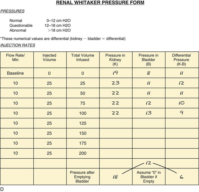
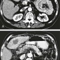
Urodynamics

Chapter 145


Urodynamics


Sally E. Mitchell


Causes of nonobstructive dilatation of the renal collecting system include vesicoureteral reflux, urinary tract infection, previous obstruction, congenital malformations, and a noncompliant bladder. Distinguishing between obstructive hydronephrosis and nonobstructive dilatation presents a problem in a number of clinical scenarios. Anatomic evaluation by means such as ultrasonography measures the degree of dilatation but does not delineate the degree of obstruction. Often, this differentiation can be made using radiologic tests such as diuretic renography and intravenous urography, as well as retrograde pyelography. However, these tests sometimes fail to conclusively determine whether an anatomic obstruction exists. It is in such cases that urodynamics can prove most useful.1


The renal collecting system and ureters are components of a dynamic system, and it is the resistance within this system that must be taken into account in determining whether an obstruction exists. Poiseuille’s equation, which originally characterized the flow of fluid through rigid tubes, has been adapted to physiologic systems. This law describes the relationship between resistance, flow, and pressure. Specifically, resistance through any conduit is directly proportional to pressure and indirectly proportionate to flow. Urodynamic studies, therefore, serve an optimal role in the evaluation of the renal collecting system and ureters because they deal directly with both the pressure and flow of fluid through these systems.


Indications


• Congenital megaureter—obstructed or nonobstructed?2,3


• Congenital megacalyces4


• Congenital ureteropelvic junction obstruction—severe enough to require surgical repair?5,6


• Any congenital hydronephrosis requiring assessment for obstruction (Fig. 145-1)


image
image
image
FIGURE 145-1 This 2-year-old boy presented with bilateral hydronephrosis. Despite bilateral nephrostomies (including two placed in different calyces on the right) and a Foley catheter, his ureters remained very dilated. To assess the ureterovesical junction for obstruction, a urinary Whitaker test was requested. A, Supine view of left collecting system during left urinary Whitaker test. Note hydronephrosis on left, dilated ureter, and trabeculated bladder. B, Supine view of right collecting system during right urinary Whitaker test. Note hydronephrosis on right, dilated and very tortuous right ureter, and more filled view of extremely trabeculated bladder. C, Pediatric urinary Whitaker test form filled out for this patient’s height (length) of 82 cm and weight of 10.9 kg. By using the DuBois chart for finding body surface area, a straight edge is placed between patient’s height and weight to cross the surface area, which can be read off the nomogram. A standard glomerular filtration rate (GFR) can be chosen by patient’s age. These numbers are then used to calculate patient’s maximal physiologic urine output per kidney in milliliters per minute. This is the rate at which urinary Whitaker test should be infused to match the rate at which kidney produces urine. In this patient, we had estimated 5 mL/min. Using the form, his rate calculated to be 4 mL/min. (In actuality, his rate of infusion should probably have been even lower because his creatinine value was 0.8 owing to chronic renal obstruction. Using the Schwartz formula, his estimated GFR was only 56.1. Using this GFR in the calculation would have calculated his maximal physiologic urine output per kidney to be 1.8 mL/min! However, keep in mind that the point of this pediatric form is to avoid infusing the adult rate of 10 mL/min in small children to avoid infusing renal pelvis above patient’s physiologic rate, which could result in collecting system rupture if there is obstruction.) Pressure results and nephroureterograms demonstrate that this patient had no ureteral obstruction but had a very trabeculated bladder with a low capacity, consistent with previously undiagnosed posterior urethral valves.

• Postoperative ureteropelvic junction repair. Is the urine draining well antegrade through the repair? This is often difficult to assess because the renal pelvis will remain large and baggy owing to the previous chronic obstruction.7,8


• Postoperative distal ureteral reimplantation. This also may be difficult to assess if the upper collecting systems remain large or dilated from previous obstruction.


• Postoperative balloon dilatation of urinary stricture


• Assessment of ureteral strictures after injury, either traumatic or surgical (Fig. 145-2)


• Assessment of malignant obstruction resolution after chemotherapy or irradiation


• Transplant kidneys with question of obstruction (Fig. 145-3)911


• Equivocal results from less invasive tests12


• Suspected obstruction in presence of poor ipsilateral renal function12


• Continued loin pain with upper urinary tract dilatation and a nonobstructive diuresis renogram12


• Unexplained loin pain that might be due to intermittent obstruction at high urine flow rates12


• Definitive investigation of the grossly dilated upper urinary tract where there is concern about the implications of an obstructive diuresis renogram with minimal symptoms12


Stay updated, free articles. Join our Telegram channel

Dec 23, 2015 | Posted by in INTERVENTIONAL RADIOLOGY | Comments Off on Urodynamics

Full access? Get Clinical Tree

Get Clinical Tree app for offline access