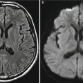
Fig. 29.1
Varicella encephalitis. (a, b) MR imaging demonstrates multiple patches and stripes of shadows with long T1 long T2 signals in the bilateral thalamus and basal ganglia, with compressed anterior horn of the right lateral ventricle. (c) By contrast imaging, the lesions are slightly enhanced. (d) CT scanning demonstrates slightly low- and even-density lesions, with unclearly defined boundaries and slightly rightward migration of the midline structure. (e, f) By a reexamination after treatment for 3 weeks, MR imaging demonstrates decreased range with lesions. (g) By DWI, the lesions located in the right basal ganglion are demonstrated in slightly high signal
29.7.2 Varicella Pneumonia
Specific X-ray demonstrations of varicella pneumonia include bilateral interstitial pneumonia and diffusive nodules. There are increased and deranged pulmonary markings in the inner and middle zone of both lungs and short fine stripes of increased density shadows in both lower lungs that interconnect into network. The nodules are obvious in the inner and middle zone of middle and lower lungs, which have a diameter of 2–10 mm, with unclearly defined boundaries. The lung apices are commonly normal. In some cases, the nodules may fuse into mass, with X-ray demonstration of confined parenchymal shadow, which is more common in adults.
In the cases of interstitial pneumonia and alveolitis, X-ray demonstrates interstitial inflammation in both lungs that is obvious in the inner and middle zone of the middle and lower lungs. There are also scattering patches of shadows or large flakes of shadows, with even density but being less obvious and unclearly defined (Figs. 29.2, 29.3, and 29.4).
Case Study 2
An infant boy, aged 6 months, with skin rashes for 4 days and fever for 2 days. The clinical diagnosis was varicella.
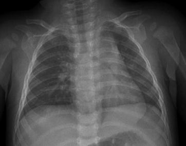
Fig. 29.2
Varicella pneumonia. X-ray radiology demonstrates small spots of blurry shadows distributing along the pulmonary markings in both lungs
Case Study 3
An infant boy, aged 5 months. He was admitted to the hospital due to fever and blister rashes for 2 days, with a body temperature of 38.1 °C (rectal temperature).
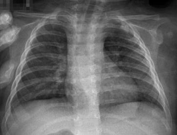
Fig. 29.3
Varicella pneumonia. X-ray demonstrates increased lung markings and multiple patches of shadows (Note: The case and images are provided by Wu, JP and Yun, J from The Third People’s Hospital of Changzhou, Changzhou, Jiangsu, China)
Case Study 4
A boy aged 8 years, with skin rashes for 10 days and fever for 4 days. He had a body temperature of 39.5 °C and was clinically diagnosed as having varicella.
Fig. 29.4
Varicella pneumonia. X-ray demonstrates patches of blurry shadows in both lungs and enlarged shadows of bilateral pulmonary hila
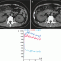
29.7.3 Severe Varicella Complicated by Pulmonary Hemorrhage
29.7.3.1 X-Ray Radiology
Chest X-ray demonstrates multiple patches and cotton-like shadows in both lungs, with lesions similar to bronchitis and pneumonia that distribute around the pulmonary markings. In some cases, the lesions diffuse outward from hilum, sharing similarities to emphysema. In addition, the transparency of the pulmonary fields decreases.
29.7.3.2 CT Scanning
In the cases with bleeding in a small quantity with a confined range, CT scanning demonstrates increased density shadows in pulmonary acinus and ground-glass-like shadows in centrilobular and panlobular areas, with interlobular septum as the interface. In the cases with bleeding in a large quantity with an extensive range, CT scanning demonstrates patches and large flakes of cloud- and cotton-like changes in diffusive ground-glass-like shadows. The lesions can be quickly and completely absorbed (Fig. 29.5).
Related posts:

Stay updated, free articles. Join our Telegram channel

Full access? Get Clinical Tree


