Vascular Anatomy
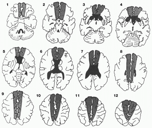 Figure 71-1. Anterior cerebral artery (ACA) territory, axial plane. The ACA territory is divided into three regions: light shading = hemispheric, medium shading = medial lenticulostriate, and dark shading = callosal. (Berman SA, Hayman LA, Hinck VC. Cerebral vascular territories: anatomic-functional correlation with axial and coronal images. In: Latchaw RE, ed. MR and CT imaging of the head, neck, and spine, 2nd ed. St. Louis: Mosby-Year Book, 1991:48-53. Figs. 3-1 and 3-2, pp. 48-49.) |
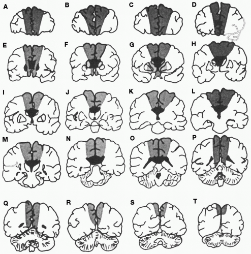 Figure 71-2. Anterior cerebral artery (ACA) territory, coronal plane. The ACA territory is divided into three regions: light shading = hemispheric, medium shading = medial lenticulostriate, and dark shading = callosal. (Berman SA, Hayman LA, Hinck VC. Cerebral vascular territories: anatomic-functional correlation with axial and coronal images. In: Latchaw RE, ed. MR and CT imaging of the head, neck, and spine, 2nd ed. St. Louis: Mosby-Year Book, 1991:48-53. Figs. 3-1 and 3-2, pp. 48-49.) |
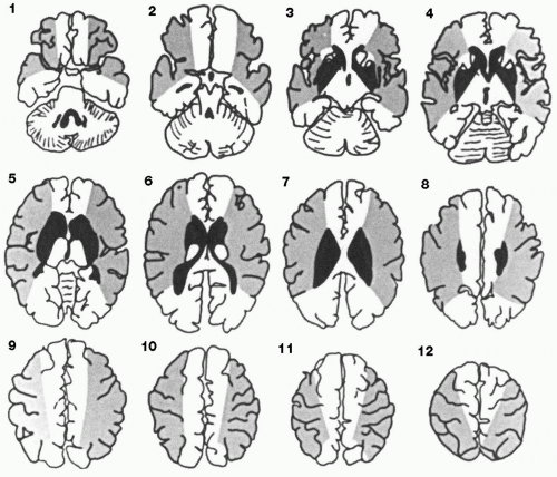 Figure 71-3. Middle cerebral artery (MCA) territory, axial plane. The MCA territory is divided into two regions: light shading = hemispheric and dark shading = lateral lenticulostriate. (Berman SA, Hayman LA, Hinck VC. Cerebral vascular territories: anatomic-functional correlation with axial and coronal images. In: Latchaw RE, ed. MR and CT imaging of the head, neck, and spine, 2nd ed. St. Louis: Mosby-Year Book, 1991:48-53. Figs. 3-4 and 3-5, pp. 50-51.) |
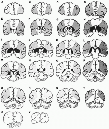 Figure 71-4. Middle cerebral artery (MCA) territory, coronal plane. The MCA territory is divided into two regions: light shading = hemispheric and dark shading = lateral lenticulostriate. (Berman SA, Hayman LA, Hinck VC. Cerebral vascular territories: anatomic-functional correlation with axial and coronal images. In: Latchaw RE, ed. MR and CT imaging of the head, neck, and spine, 2nd ed. St. Louis: Mosby-Year Book, 1991:48-53. Figs. 3-4 and 3-5, pp. 50-51.) |
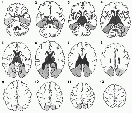 Figure 71-5. Posterior cerebral artery (PCA) territory, axial plane. The PCA territory is divided into three regions: light shading = hemispheric, medium shading = thalamic and midbrain perforators, and dark shading = callosal. (Berman SA, Hayman LA, Hinck VC. Cerebral vascular territories: anatomic-functional correlation with axial and coronal images. In: Latchaw RE, ed. MR and CT imaging of the head, neck, and spine, 2nd ed. St. Louis : Mosby-Year Book, 1991:48-53. Figs. 3-7 and 3-8, pp. 52-53.) |
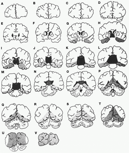 Figure 71-6. Posterior cerebral artery (PCA) territory, coronal plane. The PCA territory is divided into three regions: light shading = hemispheric, medium shading = thalamic and midbrain perforators, and dark shading
Get Clinical Tree app for offline access
Related posts:Stay updated, free articles. Join our Telegram channel
Full access? Get Clinical Tree


|