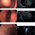
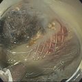
John C. Deutsch Essentia Health Care Systems, Duluth, MN, USA Vascular anomalies are often encountered incidentally during an endoscopic ultrasound (EUS) examination. They are frequently ignored as inconsequential (as in the case of atherosclerosis or calcification of the aorta). However, some vascular processes may be responsible for the symptoms which lead to the EUS examination. For example, one may find vascular occlusion of the superior mesenteric artery and celiac artery in a patient with abdominal pain, or splenic vein thrombosis in a patient with gastrointestinal (GI) hemorrhage ultimately due to isolated bleeding gastric varices. Dysphagia can be causes by aortic arch anomalies (“dysphagia lusoria”). Sometimes an unexpected vascular finding can preclude the original reason for the EUS examination as shown in a case discussed in this chapter in which an aortic pseudoaneurysm was identified during an EUS evaluation of early gastric cancer. This chapter contains images collected during EUS examinations showing a variety of vascular abnormalities that varied from the trivial to important, both symptomatic and asymptomatic. This is not intended to be comprehensive, since there are an unlimited number of conditions which could be described. Rather, this chapter will hopefully help the endosonographer to pay attention to normal and potentially abnormal vascular anatomy, since these features may be pivotal in understanding a patient’s condition. Potentially symptomatic aortic arch anomalies are relatively uncommon and occur in a small proportion (1 in 500) of the population. Those which can be easily identified during routine EUS examination include the aberrant right common carotid artery arising directly from the aortic arch. The right subclavian artery arises from the arch distal to the left subclavian artery. The right subclavian artery passes posteriorly to the trachea and esophagus. A right‐sided aortic arch can also occasionally be found. These congenital anomalies can cause swallowing problems (dysphagia lusoria). If the difficulties are severe, one can transect the artery which passes between the esophagus and the vertebrae and reimplant it. After this type of surgery a residual stump can sometimes be identified. Video 18.1 shows an example of an anomalous right subclavian artery. Video 18.2 shows a different patient in whom an anomalous right subclavian artery had been resected and reimplanted. Video 18.3 shows a right‐sided aortic arch with a vascular ring formed by an aberrant left subclavian artery, and Video 18.4 shows a different patient with the same features following surgical repair of the vascular ring. As shown in the videos, the aorta is in the right chest (to the left of the vertebrae on the image screen) through its length. At the level of the arch, the left subclavian stump can be seen. An ectatic aorta can sometimes suggest a right‐sided aorta (with the aorta in the right chest relative to the vertebrae), but this can usually be sorted out by tracing the aorta for its entire length as in Video 18.5. If the image screen on a radial array instrument is inadvertently flipped, one can also be fooled into thinking structures are on the opposite side of the chest. There are many different variants (such as double aortic arch) and branching patterns such as the bovine arch. Details concerning these varieties are available on the Internet. These features are extremely common, although the extent of disease varies. Calcification can be seen in any of the arterial structures (Video 18.6). It is usually without symptoms. The echo signal coming off the calcified structures is very bright with acoustic shadowing behind. The shadowing can interfere with visualization of perivascular structures and complicate fine needle aspiration (FNA) of small lesions. The vascular calcifications can sometimes lead to diagnostic confusion (Figure 18.1). Video 18.7 shows the computed tomography (CT) scan of a patient referred to our center for abdominal pain and chronic pancreatitis of unknown cause. Endoscopic ultrasound clearly showed that all calcifications were associated with an artery and that the pancreatic parenchyma was normal. On careful review of the CT scan, this patient appeared to have had a very redundant splenic artery and an accessory splenic artery that was the source of the abnormality seen on CT scan. The abdominal pain appeared to be unrelated to either the pancreas or the calcifications. However, vascular calcifications can sometimes be the cause of symptoms. The patient shown in Video 18.8 was referred for marked weight loss and abdominal pain suspected to be chronic pancreatitis. Esophagogastroduodenoscopy (EGD) showed what appeared to be an ischemic stomach. Endoscopic ultrasound showed a normal pancreas but obliteration of the celiac and superior mesenteric artery origins by a calcified plaque. This patient underwent urgent revascularization which corrected her pain and helped her weight return towards baseline. Plaques are not always fixed to the vessel wall. Video 18.9 shows that plaques can sometimes be noncalcified and hypermobile, suggesting a risk of embolism. An aneurysm is expansion of a vessel and all layers of the vascular wall. A pseudoaneurysm occurs when there is disruption of the vascular wall. These features can be subtle. Figure 18.2 and Video 18.10 show the area near the aortopulmonary (AP) window in a person undergoing EUS. This appears to be a pathologically enlarged lymph node. Further evaluation demonstrates this to be a pseudoaneurysm. A dramatic example of an apparent aortic pseudoaneurysm is shown in Video 18.11. This was found during evaluation of an early gastric cancer and took precedence over evaluation of the tumor. Figure 18.3 shows an example of an aortic root dissection that was first discovered during EUS for abdominal adenopathy suspected to be due to metastatic colon cancer. Figure 18.1 A radial array endoscopic ultrasound image of a calcific aortic plaque. The area behind the plaque is shadowed out. Dilation of smaller arteries is more subtle. Video 18.12 was taken from a patient in whom an outside endoscopist found a subepithelial mass and in which “the biopsies were normal.” Endoscopic ultrasound revealed a small splenic artery aneurysm. Video 18.13 shows an asymptomatic celiac artery aneurysm. In addition to degenerative atherosclerotic vascular disease, inflammatory disease such as pancreatitis can cause vascular injury as demonstrated in a patient in whom a splenic artery pseudoaneurysm was found during an exam after a resolved attack of acute pancreatitis. Video 18.14 shows a splenic artery pseudoaneurysm which arose after a bout of gallstone pancreatitis. The vessel was subsequently stented due to risk of hemorrhage. Thrombosis of the portal vein and its branches are probably the most common site for endosonographers to detect venous thrombosis. It can occur in patient with portal hypertension as shown in Figure 18.4 and Video 18.15 of a patient undergoing EUS who had cirrhosis from autoimmune liver disease. Splenic vein thrombosis can lead to isolated gastric varices. Video 18.16 shows an endoscopy from a patient referred for recurrent GI bleeding suspected to be variceal. The endoscopy shows a normal esophagus and possible gastric varicies which were confirmed during EUS. Careful evaluation of the splenic vein revealed venous obstruction from a cystic pancreatic neoplasm near the splenic hilum. Figure 18.2 A linear array image of the aortopulmonary window with an isoechoic mass that was demonstrated on the video to be a pseudoaneurysm. Figure 18.3 A linear array image showing the aortic root with clot due to an unsuspected aortic dissection. LA, left atrium; LV, left ventricle. Figure 18.4 Radial array Doppler image of a clot in the splenic vein. Dieulafoy lesions consist of arteries that are ectopically located in the submucosa. These can rupture and cause significant blood loss, but when they are not bleeding they can be very difficult to identify. Endoscopic ultrasound can be used to help identify Dieulafoy lesions as in this case (Video 18.17) in which a pulsatile lesion was seen in the rectal vault. Neoplastic tumors can extend into blood vessels and can take on the appearance of thrombus. Although rare, there are neoplastic lesions which can arise in the vascular tree and can sometimes be a source of emboli or obstruction. The most common is probably the atrial myxoma but other lesions include leiomyosarcomas and endothelial tumors. An example of a left atrial myxoma is shown in Video 18.18. Figure 18.5 shows an atrial myocardiac metastasis from breast cancer. Figure 18.6 shows a large rhabdomyosarcoma that has infiltrated into the left atrium. Figure 18.5 A linear array image of a metastasis (T) from breast cancer to the left atrial myocardium. LA, left atrium; LV, left ventricle; PA, pulmonary artery. Figure 18.6 A linear array image of a rhabdomyosarcoma infiltrating into the left atrium (LA). The mitral valve (MV) and aortic valve (AV) are shown. Arteries can be abnormally long and tortuous resulting in abnormal EUS imaging. An example is shown in Video 18.19 in which a redundant loop of splenic artery is seen. During EUS, the stomach can actually be pushed into the loop, resulting in images of the splenic artery surrounding the stomach.
18
Vascular Anomalies and Abnormalities
Introduction
Aortic arch anomalies
Vascular calcification and plaques
Aneurysms and pseudoaneurysms

Venous thrombosis


Dieulafoy lesions
Neoplasms


Miscellaneous aberrancies
Stay updated, free articles. Join our Telegram channel

Full access? Get Clinical Tree


