Abstract
Cerebral vascular anomalies are uncommon but serious abnormalities with a high risk of mortality. In this chapter, we discuss venous (aneurysm of the vein of Galen and dural sinus malformation), arterial (pial arteriovenous malformation), and mixed (hemangioma) intracranial anomalies.
Aneurysm of the vein of Galen is the most common. It can be associated both with mortality and abnormal neurodevelopment. Hyperdynamic circulation, vascular steal, and venous hypertension leading to loss of brain tissue are the mechanisms of damage. Over recent years, endovascular treatment has improved the outlook of aneurysm of the vein of Galen.
Other lesions such as arachnoid or porencephalis cysts, intracerebral hematoma, or spaces filled with cerebrospinal fluid mimic the ultrasound appearance of vascular lesions. Therefore, color flow mapping is necessary for the detection of flow. However, some types of vascular lesions may not show demonstrable flow on color flow mapping. These include hemangiomas and pial arteriovenous (AV) malformations. The use of cranial magnetic resonance imaging (MRI) can be invaluable in the assessment of cranial vascular lesions and complements ultrasound (US) use. Due to the guarded outlook of many of these lesions, pregnancy termination can be one of the options in cases with early prenatal diagnosis.
Keywords
intracranial, prenatal, vascular malformations
Introduction
Cerebral vascular malformations are rare but potentially disastrous abnormalities. Aneurysm of the vein of Galen is the most common abnormality described in prenatal series. Other types of intracranial vascular malformations are diagnosed prenatally only rarely. In the neonatal period, the clinical features include cyanosis, systolic murmur secondary to hyperdynamic circulation, cardiomegaly, and increased intracranial pressure. Before the availability of ultrasound (US) diagnosis, most cerebral vascular malformations were diagnosed only at postmortem examination. These are serious abnormalities with a high risk of mortality. The mortality rate of aneurysms of the vein of Galen is nearly 90%, with most deaths occurring within 1 week of birth. The various types of vascular malformations can be categorized as:
- •
Venous anomalies
- •
Aneurysm of the vein of Galen
- •
Dural sinus malformation (DSM)
- •
- •
Arterial anomalies
- •
Pial arteriovenous (AV) fistulas
- •
- •
Mixed anomalies
- •
Hemangiomas
- •
Aneurysm of the Vein of Galen
Definition
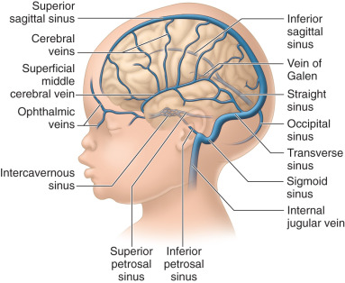
Aneurysm of the vein of Galen is a complex arteriovenous malformation (AVM) characterized by multiple communications between the system of the vein of Galen and the cerebral arteries. Fig. 42.1 is a diagrammatic representation of the cerebral venous system. This anomaly consists of dilatation of the median prosencephalic vein in association with AV fistulas within the wall of the vein, or multiple shunts communicating with the anterior part of the vein.
Prevalence and Epidemiology
Aneurysm of the vein of Galen is a rare vascular malformation; it represents less than 1% of all intracranial AVMs.
Etiology and Pathophysiology
Aneurysm of the vein of Galen is thought to be a developmental abnormality. However, several previous publications reported normal cranial US scans in the early second trimester, but a vein of Galen malformation was detected later on in the pregnancy.
Consequences of a vein of Galen malformation include the following :
- 1.
Cardiac failure: high-velocity left-to-right shunt through the AV communication causes cardiac failure. Left-to-right shunt leads to an increase in the workload of the heart, of the cardiothoracic ratio, and often of the heart rate. Postnatally, infants often have pulmonary hypertension.
- 2.
Vascular steal: AV shunt in the brain can result in a vascular steal from other arteries in the brain. The result is progressive cerebral atrophy.
- 3.
Venous hypertension: Increase in the venous pressure can lead to a reduction in the absorption of cerebrospinal fluid and progressive ventriculomegaly.
Manifestations of Disease
Imaging Technique and Findings
Ultrasound.
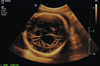
Prenatal diagnosis of the vein of Galen malformation has been made on several occasions. A classic finding is an echo-poor structure in the posterior region of the midline superior to the tentorium cerebelli and superior to the thalamus ( Fig. 42.2 ).
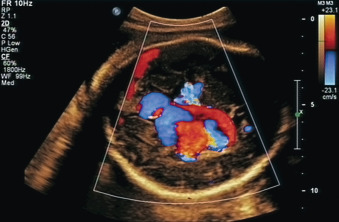
The sagittal sinus is dilated in most cases. US with color flow mapping and Doppler interrogation reveals the presence of the turbulent flow typical of an AV malformation ( Fig. 42.3 ). However, the flow may be absent if the lesion has undergone thrombosis. Three-dimensional (3D) power angiography has been used to define the structure of the malformation.
Magnetic Resonance Imaging.
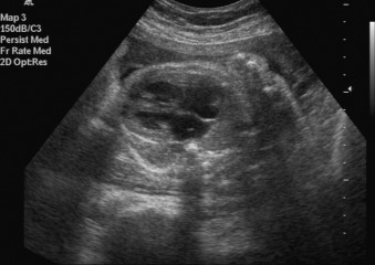
Prenatal magnetic resonance imaging (MRI) has been used to assess vascular connections and explore cerebral damage. Many associated anomalies have been described with the vein of Galen malformation. Table 42.1 lists commonly associated abnormalities. Fig. 42.4 shows cardiomegaly in a case of an aneurysm of the vein of Galen.
| Ultrasound Finding | n (%) |
|---|---|
| Cardiomegaly | 16 (64) |
| Enlarged neck veins | 8 (32) |
| Ventriculomegaly | 6 (24) |
| Polyhydramnios | 4 (16) |
| No associated findings | 6 (24) |
Synopsis of Treatment Options
Postnatal
Most affected newborns show features of cardiac failure. Control of cardiac failure is the most immediate postnatal intervention. Definitive postnatal treatment consists of total excision of the lesion, surgical ligation, or embolization. Lasjaunias et al. reported their experience of 317 cases with vein of Galen aneurysms. Initial management involved control of heart failure with diuretics and digitalis. Endovascular embolization was successfully employed as the first-line definitive treatment in 233 of 317 infants. Despite treatment, 23 (10.6%) newborns died. Severe mental retardation was reported in 20 infants, and moderate mental retardation was reported in 30 infants. Normal neurologic development was reported in 74% of survivors.
Spontaneous thrombosis of the malformation in the postnatal period has been reported. In these cases, the thrombosed lesion was seen as a mass of high density or intensity values on computed tomography (CT) or MRI. However, in most cases, the lesion does not resolve spontaneously, and definitive postnatal treatment is required.
Related posts:


Stay updated, free articles. Join our Telegram channel

Full access? Get Clinical Tree


